Review
. 2018 Apr;83(4):676-690.
doi: 10.1002/ana.25205. Epub 2018 Apr 10.

Getting the best outcomes from epilepsy surgery

Affiliations
Review

Getting the best outcomes from epilepsy surgery

Vejay N Vakharia et al. Ann Neurol. 2018 Apr.

Erratum in

  • Erratum.
    [No authors listed] [No authors listed] Ann Neurol. 2018 Jun;83(6):1205. doi: 10.1002/ana.25259. Ann Neurol. 2018. PMID: 30133830 Free PMC article. No abstract available.

Abstract

Neurosurgery is an underutilized treatment that can potentially cure drug-refractory epilepsy. Careful, multidisciplinary presurgical evaluation is vital for selecting patients and to ensure optimal outcomes. Advances in neuroimaging have improved diagnosis and guided surgical intervention. Invasive electroencephalography allows the evaluation of complex patients who would otherwise not be candidates for neurosurgery. We review the current state of the assessment and selection of patients and consider established and novel surgical procedures and associated outcome data. We aim to dispel myths that may inhibit physicians from referring and patients from considering neurosurgical intervention for drug-refractory focal epilepsies. Ann Neurol 2018;83:676-690.


Figures

Figure 1
Figure 1
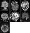
Magnetic resonance imaging of common pathologies underlying drug‐resistant focal epilepsy that are amenable to surgical treatment. (A) Coronal fluid‐attenuated inversion recovery (FLAIR) image showing increased T2 signal in the left hippocampus associated with volume loss and compensatory dilatation of the left temporal horn consistent with left hippocampal sclerosis. (B) Nonenhanced axial T1‐weighted image of a patient with a lesion in the left temporal lobe that has a “popcorn” appearance due to a hemosiderin ring and mixed intensity blood products consistent with a cavernoma. (C) Nonenhanced coronal T1‐weighted image of a patient with multiple bilateral well‐demarcated periventricular lesions that have imaging characteristics matching gray matter consistent with nodular periventricular heterotopia. This is associated with polymicrogyrialike overlying cortex. Note is also made of a posterior fossa arachnoid cyst and ventricular asymmetry. (D) Coronal FLAIR image of a patient with a sharply demarcated cortically based “pseudocystic” lesion in the right supramarginal gyrus that returns a hyperintense signal, consistent with a dysembryoplastic neuroepithelial tumor. There is associated overlying calvarial remodeling. (E) Sagittal T1‐weighted image through the left temporal lobe revealing herniation of the temporal pole through the floor of the middle cranial fossa consistent with a meningoencephalocele. (F) Coronal FLAIR image with increased signal and expansion of the left amygdala. Contrast‐enhanced imaging did not reveal any enhancement, consistent with a diffusely infiltrating low‐grade glioma. (G) Axial FLAIR image revealing increased signal in the right occipital lobe with blurring of the cortical–subcortical margin consistent with type 2B focal cortical dysplasia.
Figure 2
Figure 2
(A) Three‐dimensional cortical model. (B) Vascular segmentation from digital subtraction angiogram following left internal carotid and vertebral injections. (C) Vascular segmentation with automated parcellation of anatomical regions of interest (ROIs), including supplementary motor cortex, anterior insula, and hippocampus. (D) Automated electrode trajectory placement targeting predefined anatomical ROIs. Not all of the implemented electrode trajectories or target ROIs are shown in this image. (E) Postimplantation reconstruction of a different patient to that shown in A–D. Bolt and electrode contact points are segmented from postimplantation computed tomography and overlaid on cortical model. Electrodes with contacts implicated at seizure onset are shown in red, whereas those not involved at seizure onset are shown in white. Planned resection volume to include electrodes at seizure onset is shown in green. For clarity, the bolts are displayed, not the individual electrode contact points.
Figure 3
Figure 3
Axial, sagittal, coronal, and 3‐dimensional reconstruction of planned laser ablation trajectory (red) with outline of hippocampus (yellow), amygdala (cyan), and modeled ablation cavity (black). Other structures, such as the entorhinal cortex and parahippocampal gyrus, have been excluded for clarity. The entry point of the trajectory is centered over the crown of a gyrus, parallel to the superficial sulci. The ideal trajectory should maximize distance from vasculature, avoid crossing sulci or the lateral ventricle. In this example, the entry point is within the inferior occipital gyrus and the target point is on the anterior border of the amygdala. The Visualase (Medtronic) system is capable of performing an ablation diameter of between 5 and 20mm. The modeled ablation cavity shown above is based on a conservative estimate of 15mm.

References

    1. Engel JJ. What can we do for people with drug‐resistant epilepsy? The 2016 Wartenberg Lecture. Neurology 2016;87:2483–2489. - PMC - PubMed
    1. Wiebe S, Blume WT, Girvin JP, Eliasziw M. A randomized, controlled trial of surgery for temporal‐lobe epilepsy. N Engl J Med 2001;345:311–318. - PubMed
    1. Engel JJ, Wiebe S, French J, et al. Practice parameter: temporal lobe and localized neocortical resections for epilepsy: report of the Quality Standards Subcommittee of the American Academy of Neurology, in association with the American Epilepsy Society and the American Association of Neurology. Neurology 2003;60:538–547. - PubMed
    1. Engel JJ, McDermott MP, Wiebe S, et al. Early surgical therapy for drug‐resistant temporal lobe epilepsy. JAMA 2012;307:922–930. - PMC - PubMed
    1. Berg AT, Langfitt J, Shinnar S, et al. How long does it take for partial epilepsy to become intractable? Neurology 2003;60:186–190. - PubMed

Publication types