Review
. 2018 Mar 10;19(3):802.
doi: 10.3390/ijms19030802.

Diabetes Mellitus and Ischemic Heart Disease: The Role of Ion Channels

Affiliations
Review

Diabetes Mellitus and Ischemic Heart Disease: The Role of Ion Channels

Paolo Severino et al. Int J Mol Sci. .

Abstract

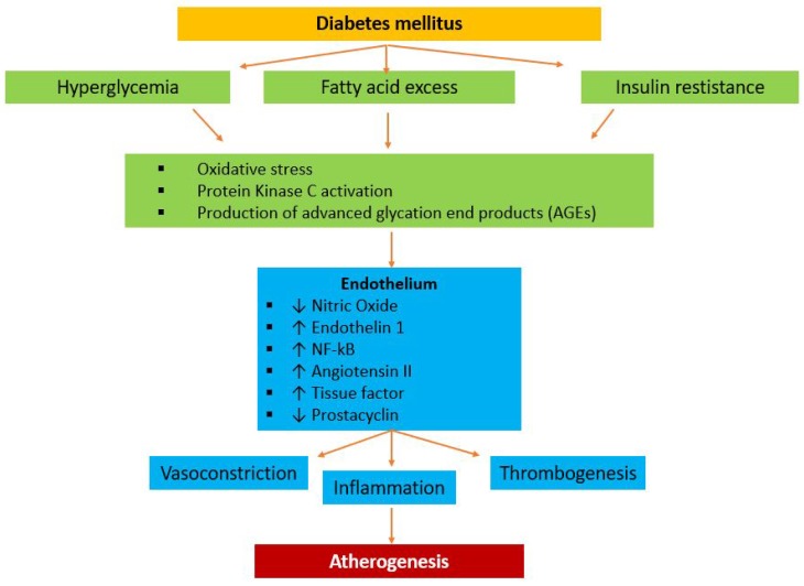
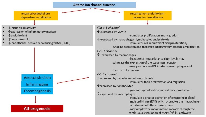
Diabetes mellitus is one the strongest risk factors for cardiovascular disease and, in particular, for ischemic heart disease (IHD). The pathophysiology of myocardial ischemia in diabetic patients is complex and not fully understood: some diabetic patients have mainly coronary stenosis obstructing blood flow to the myocardium; others present with coronary microvascular disease with an absence of plaques in the epicardial vessels. Ion channels acting in the cross-talk between the myocardial energy state and coronary blood flow may play a role in the pathophysiology of IHD in diabetic patients. In particular, some genetic variants for ATP-dependent potassium channels seem to be involved in the determinism of IHD.

Keywords: coronary blood flow; diabetes mellitus; ion channels; ischemic heart disease.


Conflict of interest statement

The authors declare no conflict of interest.

Figures

Figure 1
Figure 1
K-ATP channels and vascular tone. ATP-sensitive potassium (KATP) channels are cell membrane metabolic sensors that affect membrane excitability depending on the cellular energetic status. In normal conditions, the CK pathway is much more expressed than the AK one. A sufficient intracellular ATP concentration keeps the KATP channel closed. Hypoxia determines the activation of the AK pathway with an increase of AMP-K which opens the KATP channels. A K+ outflow causes hyperpolarization and, consequently, Ca2+ channel closing. The low intracellular Ca2+ concentration causes vasodilation. AK: Adenylate kinase ADP: Adenosine diphosphate; AMP-K: AMP-activated protein kinase; CK: Creatine kinase; PCr: Phosphocreatine.
Figure 2
Figure 2
Cl channels and vascular tone. Chloride channels are expressed in the vascular smooth muscle cells: by opening these channels, Cl ions move out of the cells and, depolarizing them, determine Ca2+ inflow with a consequent vasoconstriction effect; the opposite occurs when they are closing. Cl: Chloride.
Figure 3
Figure 3
Pathophysiology of atherogenesis in patients affected by diabetes mellitus. In diabetes, the combination of hyperglycemia, free fatty acid excess, and insulin resistance leads to several systemic effects, including increasing oxidative stress, protein kinase C, and production of advanced glycation end products. The activation of these systems impairs endothelial function, through the decreasing of nitric oxide and prostacyclin and the increasing of endothelin 1, NF-κB, angiotensin II, and tissue factor. These pathways cause vasoconstriction, increase inflammation, promote thrombosis, and, thus, contribute to atherogenesis. AGEs: advanced glycation end products.
Figure 4
Figure 4
Contribution of altered ion channel function to the pathophysiology of atherogenesis. In coronary circulation, ion channels contribute to both endothelium-dependent and non-endothelium-dependent vasodilation. When ion channel function is impaired, the vasodilating effect is decreased, affecting both pathways. In the left side of the figure (grey box), molecules and proteins involved in the impaired endothelium-dependent vasodilation are listed. These pathways are responsible of vasoconstriction, inflammation, and thrombogenesis. On the right (grey box), there is a focus on the main K channels involved in the amplification of the inflammatory cascade through the augmented proliferation and migration of lymphocytes, macrophages, and platelets. All these mechanisms contribute to atherogenesis.

Similar articles

Cited by

References

    1. World Health Organization . Global Report on Diabetes. World Health Organization; Geneva, Switzerland: 2016.
    1. Fedele F., Severino P., Bruno N., Stio R., Caira C., D’Ambrosi A., Brasolin B., Ohanyan V., Mancone M. Role of ion channels in coronary microcirculation: A review of the literature. Future Cardiol. 2013;9:897–905. doi: 10.2217/fca.13.65. - DOI - PubMed
    1. Goodwill A.G., Dick G.M., Kiel A.M. Regulation of Coronary Blood Flow. Compr. Physiol. 2017;7:321–382. doi: 10.1002/cphy.c160016. - DOI - PMC - PubMed
    1. Camici P.G., Olivotto I., Rimoldi O.E. The coronary circulation and blood flow in left ventricular hypertrophy. J. Mol. Cell. Cardiol. 2012;52:857–864. doi: 10.1016/j.yjmcc.2011.08.028. - DOI - PubMed
    1. Shapiro E. Adolf Fick–forgotten genius of cardiology. Am. J. Cardiol. 1972;30:662–665. doi: 10.1016/0002-9149(72)90606-6. - DOI - PubMed

LinkOut - more resources