

Lumbar spine epidural abscess and facet joint septic arthritis due to Streptococcus agalactiae: a case report
- PMID: 29534714
- PMCID: PMC5851089
- DOI: 10.1186/s12893-018-0350-2
Lumbar spine epidural abscess and facet joint septic arthritis due to Streptococcus agalactiae: a case report
Abstract
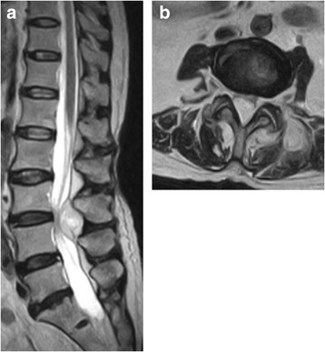
Background: Here we report a rare case of lumbar spine epidural abscess and facet joint septic arthritis caused by Streptococcus agalactiae, which had spread to the iliopsoas muscles, leading to urine retention.
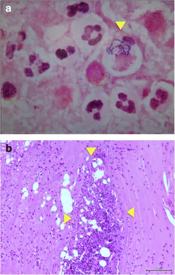
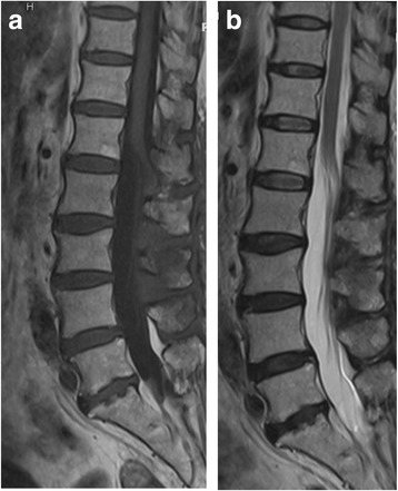
Case presentation: A 68-year-old woman with low back pain experienced a sudden onset of bilateral lower limb weakness, it was followed 14 days later by urine retention. At consultation, magnetic resonance imaging and identification of serum β-hemolytic streptococci provided a diagnosis of Streptococcus agalactiae infection. She was started on antibiotics. Despite diminishing signs of inflammation, preoperative MRI showed an epidural mass at T12-L4 compressing the cord and involving the paravertebral muscles as well. Group B beta-hemolytic streptococci were detected in both urine and blood. Because of bilateral lower limb weakness and urine retention, T12-L4 hemilaminectomy was performed. The L3/L4 intertransverse ligament resected and abscess drained. Histopathology revealed that inflammatory cells had invaded the facet joint. Group B beta-hemolytic streptococci were identified, confirming the diagnosis. The patient continued with the antibiotics postoperatively, and her health rapidly improved.
Conclusion: Lumbar spine epidural abscess and facet joint septic arthritis caused by Streptococcus agalactiae is a clinical emergency, with significant morbidity and mortality especially with delayed diagnosis. A delay in both diagnosis and aggressive treatment can lead to not only severe neurological deficit but also to septicaemia, multiorgan failure, and even death.
Keywords: Antibiotic administration; Facet joint septic arthritis; Hemilaminectomy; Spinal epidural abscess; Streptococcus agalactiae; Urine retention.
Conflict of interest statement
Ethics approval and consent to participate
Not applicable.
Consent for publication
Written informed consent was obtained from the patient for publication of this case report and accompanying images.
Competing interests
The authors declare that they have no competing interests.
Publisher’s Note
Springer Nature remains neutral with regard to jurisdictional claims in published maps and institutional affiliations.
Figures


References
-
- Chung S, Chen C, Yu W. Spinal epidural abscess caused by group B Streptococcus in a diabetic woman presenting with febrile low back pain. Jpn J Infect Dis. 2005;58(3):177. - PubMed
Publication types
MeSH terms
Substances
LinkOut - more resources
Full Text Sources
Other Literature Sources
Medical
