. 2018 Mar 13;16(1):10.
doi: 10.1186/s12964-018-0223-4.

Ras enhances TGF-β signaling by decreasing cellular protein levels of its type II receptor negative regulator SPSB1

Affiliations

Ras enhances TGF-β signaling by decreasing cellular protein levels of its type II receptor negative regulator SPSB1

Sheng Liu et al. Cell Commun Signal. .

Abstract

Background: Transformation by oncogene Ras overcomes TGF-β mediated growth inhibition in epithelial cells. However, it cooperates with each other to mediate epithelial to mesenchymal transition (EMT). The mechanism of how these two pathways interact with each other is controversial.

Methods: Molecular techniques were used to engineer expression plasmids for Ras, SPRY, TGF-β receptors, type I and II and ubiquitin. Immunoprecipitation and western blots were employed to determine protein-protein interactions, preotein levels, protein phosphorylation while immunofluorecesent staining for molecular co-localization. TGF-β signalling activities is also determined by its luciferase reporter assay. Trans-well assays were used to measure cell migration and invasion.

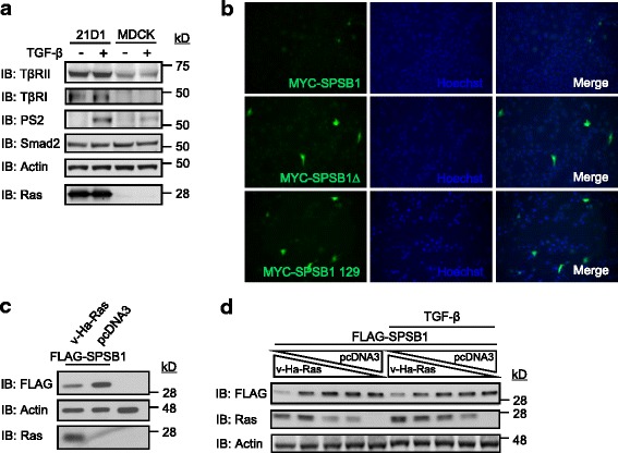
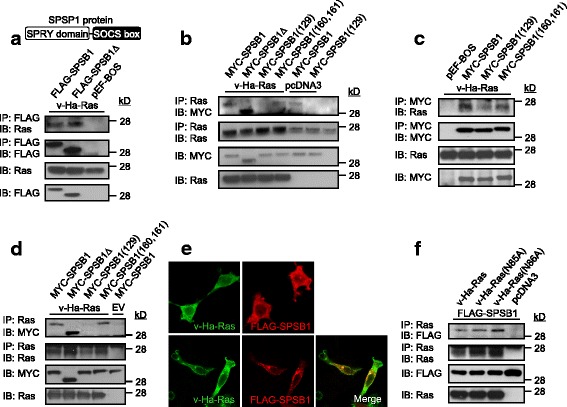
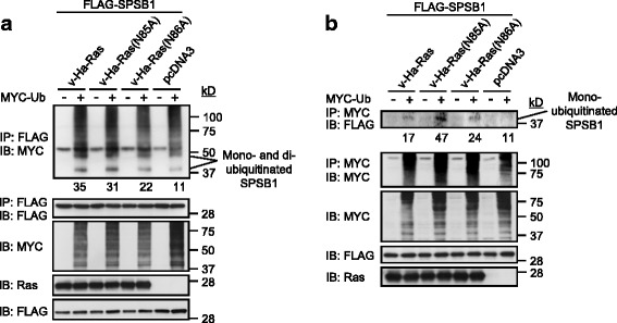
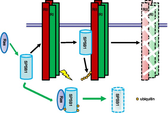
Results: Ras interacts with the SPSB1's SPRY domain to enhance TGF-β signaling. Ras interacts and colocalizes with the TGF-β type II receptor's (TβRII) negative regulator SPSB1 on the cell membrane, consequently promoting SPSB1 protein degradation via enhanced mono- and di-ubiquitination. Reduced SPSB1 levels result in the stablization of TβRII, in turn the increase of receptor levels significantly enhance Smad2/3 phosphorylation and signaling. Importantly, forced expression of SPSB1 in Ras transformed cells suppresses TGF-β signaling and its mediated migration and invasion.

Conclusion: Ras positively cooperates with TGF-β signaling by reducing the cellular protein levels of TβRII negative regualtor SPSB1.

Keywords: Ras; SPSB1; TGF-β signaling.


Conflict of interest statement

Ethics approval and consent to participate

Gene manipulations were conducted in according with University of Melbourne Gene Technology and Biosafety Committee (IBC No 301) approval 2014/008.

Consent for publication

All authors consent for publication.

Competing interests

The authors declare that they have no competing interests.

Publisher’s Note

Springer Nature remains neutral with regard to jurisdictional claims in published maps and institutional affiliations.

Figures

Fig. 1
Fig. 1
Oncogenic Ras reduces SPSB1 expression level. Cultured MDCK and 21D1 cells (a) were treated ± TGF-β (2 ng/ml) for 15 min and then lysed. Whole cell lysates were examined for indicated proteins by immunoblotting (IB). 21D1 cells (b) were transfected with indicated DNA constructs (0.2 μg/well each) in a 12 well plate for 48 h. Fixed cells were then immunostained with mouse anti-MYC followed by Alexa488-conjugated secondary anti-mouse IgG. Cell nuclei was stained with Hoechst dye. The expression of SPSB1/SPSB1 mutants (green) was analyzed by fluorescent microscope (magnification = 40×). 293 T cells (c) were transfected with indicated DNA constructs for 48 h. Cells in (d) were transfected with the FLAG-SPSB1 (0.5 μg/well) and decreasing concentration of v-Ha-Ras (0.5, 0.25, 0.125, 0.0625, 0 μg/well), the total amount of DNA per transfection was kept the same by compensating with pcDNA3 vector. 24 h later, cells were treated with ± TGF-β (2 ng/ml) for a further 24 h. Whole cell lysates were examined for indicated proteins by immunoblotting (IB). In all case, each experiment was repeated, with representative results shown
Fig. 2
Fig. 2
Ras interacts with the SPRY domain of SPSB1. 293 T cells (a, b, c, d, f) were transfected with indicated DNA constructs (0.5 μg/well each). 48 h later, cell lysates were immunoprecipitated (IP) with anti-Ras antibody (b, d, f) or anti-MYC antibody (c) conjugated with protein G beads or anti-FLAG beads (a). Both whole cell lysates and immunoprecipatates were examined for indicated proteins by immunoblotting (IB). 293 T cells (e) were transfected with v-Ha-Ras or FLAG-SPSB1 singly (top two images) or co-transfected with v-Ha-Ras and FLAG-SPSB1 (bottom images) for 48 h. Fixed cells were then immunostained with rabbit anti-FLAG followed by Alexa546-conjugated secondary anti-rabbit IgG and/or mouse anti-Ras followed by Alexa488-conjugated secondary anti-mouse IgG as indicated. The sub-cellular localization of SPSB1 (red) and v-Ha-Ras (green) was analyzed by confocal microscope (magnification = 60×). Co-localization of merged images appears as yellow. All experiments were repeated three times, with representative results shown. A figure illustration of SPSB1 protein is shown in a
Fig. 3
Fig. 3
Oncogenic Ras enhances the ubiquitination levels of SPSB1. 293 T cells (a, b) were transfected with indicated FLAG-SPSB1 construct together with v-Ha-Ras/v-Ha-Ras mutants or pcDNA3 control vector ± MYC-ubiquitin. Cells were lysed 48 h post-transfection. Cell lysates were immunoprecipitated (IP) with FLAG beads (a) or anti-MYC antibody conjugated with protein G beads (b). Both whole cell lysates and immunoprecipatates were examined for indicated proteins by immunoblotting (IB). Relative densities of mono-ubiquitinated bands are list below the top panels. Results are representative of experiments repeated at least once
Fig. 4
Fig. 4
Oncogenic Ras increases the degradation rate of SPSB1. 293 T cells (a, b, c, d) were co-transfected with various SPSB1 and Ras/control construct as indicated. 36 h later, cells were exposed to cycloheximide (20 μg/ml) for indicated periods. Cells in (d) were co-treated with MG132 (25 μM) for indicated periods. Whole cell lysates were then examined for indicated proteins by immunoblotting (IB). Relative intensity of each SPSB1/SPSB1 mutant band (a and c) was qualitatively measured using ImageJ, the number under each band indicated its corresponding relative intensity (Arbitrary Units). The half life ± S.D. (n = 3 technical replicates) for SPSB1/SPSB1 mutant degradation is shown underneath. In all case, each experiment was repeated at least once, one representing result is showing
Fig. 5
Fig. 5
Oncogenic Ras reduces the enhanced ubiquitination of TβRII by SPSB1 and hence stabilizes TβRII. 293 T cells (a, b) were transfected with indicated FLAG-SPSB1/pEF-BOS control vector together with v-Ha-Ras/pcDNA3 control vector and HA-TβRII ± MYC-ubiquitin. Cells (a) were lysed 48 h post-transfection. Cell lysates were immunoprecipitated (IP) with anti-TβRII antibody conjugated with protein G beads. 36 h post-transfection, cells (b) were exposed to cycloheximide (20 μg/ml) for indicated periods and lysed. In all cases, both whole cell lysates and immunoprecipatates were examined for indicated proteins by immunoblotting (IB). Relative intensity of each TβRII band (b) was qualitatively measured using ImageJ as described in Fig. 4. In all case, results are representative of experiments repeated at least once
Fig. 6
Fig. 6
TGF-β receptor levels regulate TGF-β signaling sensitivity and duration. 293 T cells (a) were co-transfected with pCAGA-luc and indicated TβRII and/or TβRI and/or pcDNA3 control vector. 24 h later, cells were treated with ± TGF-β at indicated concentration for a further 24 h. Cells were then lysed, and luciferase activity was determined. Data are expressed as relative Smad3 luciferase activity (fold of induction) by standardizing the luciferase activity of un-stimulated cells transfected with contorl vector to 1, and normalizing all other raw values accordingly. Results from a representative experiment are shown as the mean of triplicates±S.D. * P < 0.05. 293 T cells (b) were co-transfected with indicated DNA constructs. 36 h later, cells were stimulated with ± TGF-β (2 ng/ml) for indicated periods then lysed. Whole cell lysates were examined for indicated proteins by immunoblotting (IB). In all case, each experiment was repeated three times, one representing result is shown
Fig. 7
Fig. 7
Oncogenic Ras enhances TGF-β signaling. Doxycycline inducible v-Ha-Ras MDCK cells (a) were cultured in ± doxycycline (2 μg/ml) for 2 weeks. Cells were then transfected with pCAGA-luc. MDCK and 21D1 cells (b) were transfected with pCAGA-luc. 293 T cells (c, d) were co-transfected with pCAGA-luc and indicated combination of TβRII, TβRI, SPSB1, Ras, pcDNA3 and pEF-BOS constructs. In all cases, 24 h post-transfection, cells were treated with ± TGF-β (2 ng/ml in a) at indicated concentration for a further 24 h and lysed. Luciferase activity was determined as desribed in Fig. 6. Data are expressed as mean relative Smad3 luciferase activity (fold-induction) and error bars represent S.D. from representative experiments performed 3 times. * P < 0.05. Western blot of the expression levels of v-Ha-Ras (a) was conducted using the same cell lysates for luciferasμe assay. Results are representative of experiments repeated at least once
Fig. 8
Fig. 8
Reducing TGF-β signaling in oncogenic Ras transformed MDCK cells by SPSB1 suppresses cell migration and invasion. 21D1 cells (a and c) were co-transfected with eGFP construct and FLAG-SPSB1 or pEF-BOS for 48 h. Cell monolayers (a) were then scratched as described in materials and methods, treated with ± TGF-β (2 ng/ml) and phase contrast/fluorescence images were recorded at 0 and 24 h post-scratching. Total cell number (denominator) and eGFP expressing cell number (numerator) that migrated into the wounded area were counted and represented as relative cell migration in B. Similar results were obtained in three independent experiments. c Following DNA transfection, cell were collected and re-seeded into the upper chamber of 8 μM pore matrigel-coated transwell plates (matrigel 1:1 mixed with DMEM, 70 μl/well) ± TGF-β (2 ng/ml) as indicated for another 24 h. Cells that migrated to the bottom side of the upper chamber were then fixed and stained with Hoechst dye. Images were taken using a fluorescence microscope (magnification = 20×) in 4 random fields. Total cell number (denominator) and eGFP expressing cell number (numerator) that migrated to the bottom side of the upper chamber were counted and represented as relative cell migration. Bars represent the mean ± S.D. of triplicate wells from one of three representative experiments
Fig. 9
Fig. 9
Schematic illustration of Ras enhancing TGF-β signaling through decreasing of SPSB1, a TβRII ubiquitination regulator. SPSB1 interacts with TβRII/TβRI (RII/RI), resulting in polyubiquitination and degradation of the type II receptor (black arrows). Ras interacts with SPSB1, resulting in its protein level decrease through mono-, di-ubiquitination of SPSB1 (green arrows). Consequentluy, Ras enhances TGF-β signaling

Similar articles

Cited by

References

    1. Zhu HJ, Burgess AW. Regulation of transforming growth factor-beta signaling. Mol Cell Biol Res Commun. 2001;4:321–330. doi: 10.1006/mcbr.2001.0301. - DOI - PubMed
    1. Markowitz S, Wang J, Myeroff L, Parsons R, Sun L, Lutterbaugh J, Fan RS, Zborowska E, Kinzler KW, Vogelstein B, et al. Inactivation of the type II TGF-beta receptor in colon cancer cells with microsatellite instability. Science. 1995;268:1336–1338. doi: 10.1126/science.7761852. - DOI - PubMed
    1. Hahn SA, Schutte M, Hoque AT, Moskaluk CA, da Costa LT, Rozenblum E, Weinstein CL, Fischer A, Yeo CJ, Hruban RH, Kern SE. DPC4, a candidate tumor suppressor gene at human chromosome 18q21.1. Science. 1996;271:350–353. doi: 10.1126/science.271.5247.350. - DOI - PubMed
    1. Das P, Maduzia LL, Padgett RW. Genetic approaches to TGFbeta signaling pathways. Cytokine Growth Factor Rev. 1999;10:179–186. doi: 10.1016/S1359-6101(99)00014-3. - DOI - PubMed
    1. Akhurst RJ, Derynck R. TGF-beta signaling in cancer--a double-edged sword. Trends Cell Biol. 2001;11:S44–S51. - PubMed

Publication types

MeSH terms

Substances

LinkOut - more resources