. 2018 Mar 13;15(1):76.
doi: 10.1186/s12974-018-1110-z.

Teriflunomide promotes oligodendroglial differentiation and myelination

Affiliations

Teriflunomide promotes oligodendroglial differentiation and myelination

Peter Göttle et al. J Neuroinflammation. .

Abstract

Background: Multiple sclerosis (MS) is a neuroinflammatory autoimmune disease of the central nervous system (CNS) which in most cases initially presents with episodes of transient functional deficits (relapsing-remitting MS; RRMS) and eventually develops into a secondary progressive form (SPMS). Aside from neuroimmunological activities, MS is also characterized by neurodegenerative and regenerative processes. The latter involve the restoration of myelin sheaths-electrically insulating structures which are the primary targets of autoimmune attacks. Spontaneous endogenous remyelination takes place even in the adult CNS and is primarily mediated by activation, recruitment, and differentiation of resident oligodendroglial precursor cells (OPCs). However, the overall efficiency of remyelination is limited and further declines with disease duration and progression. From a therapeutic standpoint, it is therefore key to understand how oligodendroglial maturation can be modulated pharmacologically. Teriflunomide has been approved as a first-line treatment for RRMS in the USA and the European Union. As the active metabolite of leflunomide, an established disease-modifying anti-rheumatic drug, it mainly acts via an inhibition of de novo pyrimidine synthesis exerting a cytostatic effect on proliferating B and T cells.

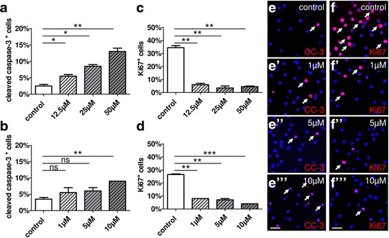
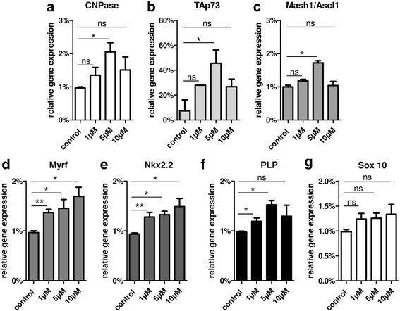
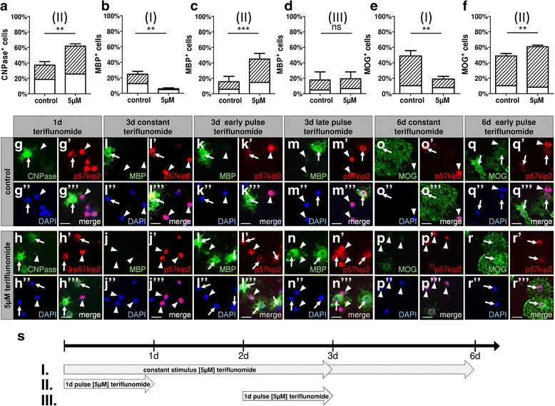
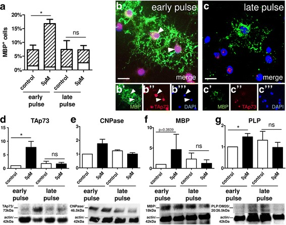
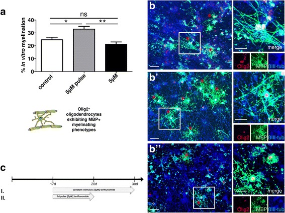
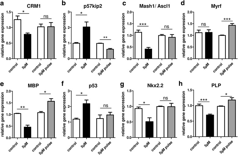
Methods: We investigated teriflunomide-dependent effects on primary rat oligodendroglial homeostasis, proliferation, and differentiation related to cellular processes important for myelin repair hence CNS regeneration in vitro. To this end, several cellular parameters, including specific oligodendroglial maturation markers, in vitro myelination, and p53 family member signaling, were examined by means of gene/protein expression analyses. The rate of myelination was determined using neuron-oligodendrocyte co-cultures.

Results: Low teriflunomide concentrations resulted in cell cycle exit while higher doses led to decreased cell survival. Short-term teriflunomide pulses can efficiently promote oligodendroglial cell differentiation suggesting that young, immature cells could benefit from such stimulation. In vitro myelination can be boosted by means of an early stimulation window with teriflunomide. p73 signaling is functionally involved in promoting OPC differentiation and myelination.

Conclusion: Our findings indicate a critical window of opportunity during which regenerative oligodendroglial activities including myelination of CNS axons can be stimulated by teriflunomide.

Keywords: Inhibitor; Multiple sclerosis; Myelin repair; Neuroregeneration; Oligodendrocyte; Remyelination; Transcription factor; p57kip2; p73.


Conflict of interest statement

Ethics approval

Experimental procedures were approved by the Institutional Animal Care and Use Committee of the Heinrich Heine University in accordance with the criteria outlined in the Institutional Guidelines for Animal Research.

Consent for publication

Not applicable.

Competing interests

AM and LR have no competing interests. PG and PK performed consultancy work for Geneuro. DK received compensation for speaking from Grifols SA. HPH received compensation for consulting, speaking, and serving on steering committees from Bayer Healthcare, Biogen, Geneuro, MedImmune, Merck, Novartis, Opexa, Receptos Celgene, Roche, Sanofi Genzyme, and Teva with the approval by the Rector of Heinrich Heine University.

Publisher’s Note

Springer Nature remains neutral with regard to jurisdictional claims in published maps and institutional affiliations.

Figures

Fig. 1
Fig. 1
Oligodendroglial cell death and proliferation rates upon stimulation with teriflunomide. a Application of high doses of teriflunomide for 1 day (1d) led to a substantial increase in the percentage of apoptotic OPCs cells as judged by the expression of activated caspase-3 (CC-3), whereas low concentration profiles (b, ee”’) did not significantly change the naturally occurring death rate. Proliferation after 1d was significantly reduced upon high (c) and low (d, ff”’) doses as indicated by the percentage of Ki-67-positive cells. Data are shown as mean; error bars represent SEM. Number of experiments: n = 2 for (ad). t test (ns, not significant, *p < 0.05, **p < 0.01, ***p < 0.001). Scale bars, 30 μM
Fig. 2
Fig. 2
ag Gene expression responses upon short-term teriflunomide stimulation. Quantitative RT-PCR after 1d revealed that stimulation of primary rat OPCs with tolerated concentration profiles of teriflunomide led to a significant upregulation of CNPase, TAp73, Mash1/Ascl1, Myrf, Nkx2.2, PLP, and Sox 10 transcript levels. GAPDH was used as reference gene. Data are shown as mean, and error bars represent SEM. Number of experiments: n = 3. t test (ns, not significant, *p < 0.05, **p < 0.01)
Fig. 3
Fig. 3
OPC differentiation dynamics and marker protein responses as a function of different teriflunomide application schemes. af Percentage of oligodendroglial cells positive for myelin markers displaying nuclear (white bars) or cytoplasmic (dashed bars) p57kip2 signals. a, gh”’ After short-term stimulation with 5 μM teriflunomide (s; scheme II), an increase of CNPase-positive cells was observed along with increased translocation of p57kip2 from the nucleus (arrowheads) to the cytoplasm (arrows). c, kl”’ A 24 h pulse stimulation (scheme II) with teriflunomide followed by a 48-h (or 120-h; f, qr”’) withdrawal period led to a substantial increase in the fraction of oligodendroglial cells that translocated p57kip2 from the nucleus to the cytoplasm and correlated with an increase of MBP- and MOG-positive cells, respectively. b, ij”’ On the other hand, long-term stimulation (scheme I) over 72 h (or 144 h; e, op”’) resulted in a decrease of cells expressing MBP or MOG and boosted nuclear accumulation of the p57kip2 protein. d, mn”’ Moreover, a short-term pulse at a later time point from 48 to 72 h (scheme III) did not affect oligodendroglial differentiation. Data are shown as mean, and error bars represent SEM. Number of experiments: n = 3 (af). t test (ns, not significant, **p < 0.01, ***p < 0.001). Scale bars, 20 μM
Fig. 4
Fig. 4
p73 protein induction in response to teriflunomide stimulation. ac”’ Double immunostaining and its quantification confirmed that early teriflunomide pulses (scheme II, see Fig. 3) result in cells displaying strong TAp73 signals (arrowheads in b) correlating also with MBP positivity (dashed bars in a). Late teriflunomide pulses (scheme III) could not boost both protein markers. Scale bars, 20 μm. dg Western blot analysis confirmed that early short-term teriflunomide pulses (scheme II) result in a strong induction of CNPase (e), MBP (f), and PLP levels (g), as well as of TAp73 (d) protein levels. Late pulses (scheme III) did not upregulate TAp73 or myelin marker expression (as quantified for TAp73 in d). Actin was used for normalization and protein molecular weights are indicated in kilodalton. Data are shown as mean, and error bars represent SEM. t test (ns, not significant, *p < 0.05). Number of experiments: n = 3 for (a, d, f, g), n = 2 for (e)
Fig. 5
Fig. 5
Teriflunomide-dependent modulation of myelination in vitro. Myelinating neuron/oligodendrocyte co-cultures were fixed and stained after 30 days in vitro (DIV30). a Early short-term (3d) teriflunomide pulse stimulation (scheme II; gray bar; b’) significantly increased the number of Olig2-positive oligodendrocytes that formed MBP-positive internodes (asterisk) as compared to controls (white bar; b) or long-term stimulation (scheme I; black bar; b”). bb” Representative triple staining for Olig2, MBP, and ßIII-tubulin. Scale bars, 30 μm. Data are shown as mean, and error bars represent SEM. t test (ns, not significant, *p < 0.05, **p < 0.01). Number of experiments: n = 5 for (a)
Fig. 6
Fig. 6
Differential gene expression responses upon constant versus pulsed teriflunomide stimulation. Quantitative RT-PCR upon constant teriflunomide stimulation (scheme I, Fig. 3; black bars) revealed a significant decrease in transcript levels of CRM1 (a), MBP (e), PLP (h), and of transcription factors Mash1/Ascl1 (c) and Nkx2.2 (g), whereas expression of the oligodendroglial differentiation inhibitor p57kip2 (b) as well as of the stress response protein p53 (f) were increased. Short-term teriflunomide pulse stimulation (scheme II; gray bars) exerted no significant impact on the expression of CRM1, Mash1/Ascl1, Nkx2.2, and p53 but significantly reduced the expression of the OPC differentiation inhibitor p57kip2 and induced Myrf (d), MBP, and PLP transcript levels. GAPDH and ODC were used as reference genes. Data are shown as mean, and error bars represent SEM. t test (ns, not significant, *p < 0.05, **p < 0.01, ***p < 0.001). Number of experiments: n = 3 for (ah)

Similar articles

Cited by

References

    1. Kutzelnigg A, Lassmann H. Pathology of multiple sclerosis and related inflammatory demyelinating diseases. Handb Clin Neurol. 2014;122:15–58. doi: 10.1016/B978-0-444-52001-2.00002-9. - DOI - PubMed
    1. Trapp BD, Peterson J, Ransohoff RM, Rudick R, Mork S, Bo L. Axonal transection in the lesions of multiple sclerosis. N Engl J Med. 1998;338:278–285. doi: 10.1056/NEJM199801293380502. - DOI - PubMed
    1. Franklin RJ, Ffrench-Constant C, Edgar JM, Smith KJ. Neuroprotection and repair in multiple sclerosis. Nat Rev Neurol. 2012;8:624–634. doi: 10.1038/nrneurol.2012.200. - DOI - PubMed
    1. Claussen MC, Korn T. Immune mechanisms of new therapeutic strategies in MS: teriflunomide. Clin Immunol. 2012;142:49–56. doi: 10.1016/j.clim.2011.02.011. - DOI - PubMed
    1. Bruneau JM, Yea CM, Spinella-Jaegle S, Fudali C, Woodward K, Robson PA, Sautes C, Westwood R, Kuo EA, Williamson RA, Ruuth E. Purification of human dihydro-orotate dehydrogenase and its inhibition by A77 1726, the active metabolite of leflunomide. Biochem J. 1998;336(Pt 2):299–303. doi: 10.1042/bj3360299. - DOI - PMC - PubMed

MeSH terms