

Editorial
doi: 10.1681/ASN.2018020200.
Epub 2018 Mar 13.
Maintenance and Breakdown of Glomerular Tuft Architecture
Affiliations
- PMID: 29535166
- PMCID: PMC5875972
- DOI: 10.1681/ASN.2018020200
Item in Clipboard
Editorial
Maintenance and Breakdown of Glomerular Tuft Architecture
J Am Soc Nephrol.
2018 Apr.
No abstract available
Keywords: GBM; adhesion complex; mesangial cell.
Figures
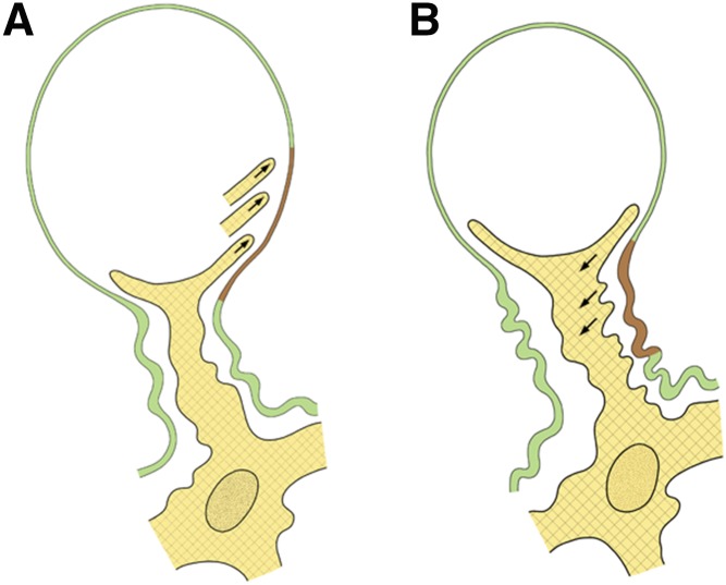
Mechanisms of changing the luminal width of glomerular capillaries shown in schematics. (A) Decreasing the capillary lumen (from A to B). Mesangial cell processes extend into the space between the endothelium and the glomerular basement membrane (GBM; arrows) and establish contacts to more peripheral sites of the GBM. By contraction, peripheral parts of the GBM (shown in brown) are pulled centripetally, and as seen in B, they are added to the paramesangial GBM. (B) Increasing the capillary lumen (from B to A). Mesangial cell processes disconnect from the GBM and retract (arrows). Thereby, the most peripheral portions of the paramesangial GBM (shown in brown) are released from the mesangium, and as seen in A, they are added to the peripheral portion leading to capillary expansion driven by the pressure gradient.
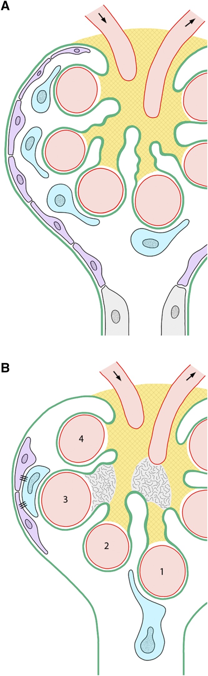
Consequences of mesangial failures for podocytes shown in schematics. (A) Normal situation. The integrity of the mesangium is indicated by a checker pattern. Podocytes are shown in blue. Parietal epithelial cells are shown in violet. (B) A breakdown of mesangial cell-glomerular basement membrane connections is shown at two sites indicated by the disappearance of the checker pattern. The consequences with respect to capillary 1 consist of a lengthening of the corresponding mesangial axis followed by the prolapse of the capillary and the associated podocyte into the urinary orifice., With respect to capillary 3, the consequences of a breakdown of mesangial cell-glomerular basement membrane connections consist of a bulging of the mesangium and an expansion of the capillary followed by the peripheral displacement of the associated podocytes, which form contacts to the parietal epithelium. Correspondingly, displacement of capillary 2 would be followed by a tip lesion of capillary 4 by a vascular pole–associated tuft adhesion.
Comment on
-
Nephronectin Regulates Mesangial Cell Adhesion and Behavior in Glomeruli.J Am Soc Nephrol. 2018 Apr;29(4):1128-1140. doi: 10.1681/ASN.2017070752. Epub 2018 Jan 15. J Am Soc Nephrol. 2018. PMID: 29335243 Free PMC article.
References
-
- Sakai T, Kriz W: The structural relationship between mesangial cells and basement membrane of the renal glomerulus. Anat Embryol (Berl) 176: 373–386, 1987 - PubMed
-
- Elger M, Sakai T, Kriz W: Role of mesangial cell contraction in adaptation of the glomerular tuft to changes in extracellular volume. Pflugers Arch 415: 598–605, 1990 - PubMed
Publication types
MeSH terms
Substances
LinkOut - more resources
Full Text Sources
Other Literature Sources
