. 2018 Mar 13;9(2):e00246-18.
doi: 10.1128/mBio.00246-18.

Serum High-Mobility-Group Box 1 as a Biomarker and a Therapeutic Target during Respiratory Virus Infections

Affiliations

Serum High-Mobility-Group Box 1 as a Biomarker and a Therapeutic Target during Respiratory Virus Infections

Mira C Patel et al. mBio. .

Abstract

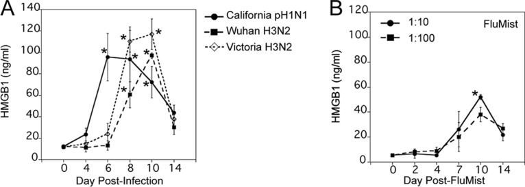
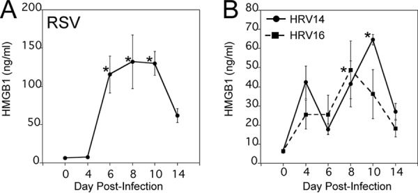
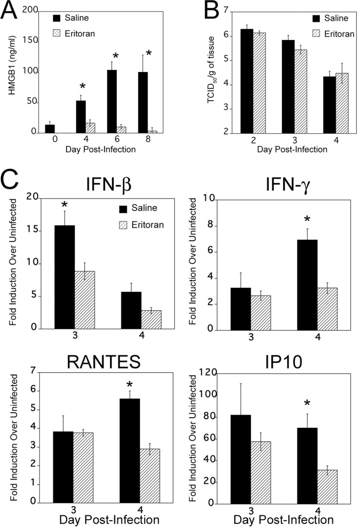
Host-derived "danger-associated molecular patterns" (DAMPs) contribute to innate immune responses and serve as markers of disease progression and severity for inflammatory and infectious diseases. There is accumulating evidence that generation of DAMPs such as oxidized phospholipids and high-mobility-group box 1 (HMGB1) during influenza virus infection leads to acute lung injury (ALI). Treatment of influenza virus-infected mice and cotton rats with the Toll-like receptor 4 (TLR4) antagonist Eritoran blocked DAMP accumulation and ameliorated influenza virus-induced ALI. However, changes in systemic HMGB1 kinetics during the course of influenza virus infection in animal models and humans have yet to establish an association of HMGB1 release with influenza virus infection. To this end, we used the cotton rat model that is permissive to nonadapted strains of influenza A and B viruses, respiratory syncytial virus (RSV), and human rhinoviruses (HRVs). Serum HMGB1 levels were measured by an enzyme-linked immunosorbent assay (ELISA) prior to infection until day 14 or 18 post-infection. Infection with either influenza A or B virus resulted in a robust increase in serum HMGB1 levels that decreased by days 14 to 18. Inoculation with the live attenuated vaccine FluMist resulted in HMGB1 levels that were significantly lower than those with infection with live influenza viruses. RSV and HRVs showed profiles of serum HMGB1 induction that were consistent with their replication and degree of lung pathology in cotton rats. We further showed that therapeutic treatment with Eritoran of cotton rats infected with influenza B virus significantly blunted serum HMGB1 levels and improved lung pathology, without inhibiting virus replication. These findings support the use of drugs that block HMGB1 to combat influenza virus-induced ALI.IMPORTANCE Influenza virus is a common infectious agent causing serious seasonal epidemics, and there is urgent need to develop an alternative treatment modality for influenza virus infection. Recently, host-derived DAMPs, such as oxidized phospholipids and HMGB1, were shown to be generated during influenza virus infection and cause ALI. To establish a clear link between influenza virus infection and HMGB1 as a biomarker, we have systematically analyzed temporal patterns of serum HMGB1 release in cotton rats infected with nonadapted strains of influenza A and B viruses and compared these patterns with a live attenuated influenza vaccine and infection by other respiratory viruses. Towards development of a new therapeutic modality, we show herein that blocking serum HMGB1 levels by Eritoran improves lung pathology in influenza B virus-infected cotton rats. Our study is the first report of systemic HMGB1 as a potential biomarker of severity in respiratory virus infections and confirms that drugs that block virus-induced HMGB1 ameliorate ALI.

Keywords: HMGB1; biomarker; influenza; respiratory syncytial virus; respiratory viruses.


Figures

FIG 1
FIG 1
Increased serum HMGB1 levels in cotton rats infected with influenza B/Wisconsin virus. (A) Young male cotton rats were infected i.n. with 1 × 10⁶ TCID50/rat of influenza B/Wisconsin virus (Live Influenza B). Another group of animals similarly inoculated with UV-inactivated influenza B/Wisconsin virus (UV Influenza B) (at a dose similar to that of live virus) were used as controls. Serum samples were obtained on the indicated days p.i., and HMGB1 levels were analyzed by ELISA. Results are expressed as geometric means ± standard errors of the means (SEMs) (error bars) of the quantity of HMGB1 (in nanograms per milliliter) at each indicated time. Values for the group inoculated with live influenza virus that were significantly different (P < 0.05) from the values for the group inoculated with UV-inactivated influenza B virus are indicated by an asterisk. There were five cotton rats in each group for each treatment and time point. (B) Age-matched male and female cotton rats were infected i.n. with 1 × 10⁶ TCID50 of live influenza B virus per rat. Serum samples were obtained and analyzed for HMGB1 levels at the indicated days p.i. There were five cotton rats in each group at each time point. (C) Groups (five cotton rats in each group) were infected i.n. with either 1 × 10⁵ or 1 × 10⁶ TCID50 of live influenza B virus per rat, and serum HMGB1 levels were analyzed at the indicated days p.i. The values for rats infected with 1 × 10⁶ TCID50 that were significantly different (P < 0.05) from the values for rats infected with 1 × 10⁵ TCID50 are indicated by an asterisk.
FIG 2
FIG 2
Serum HMGB1 levels increase upon infection with various strains of human influenza A viruses in cotton rats. (A) Cotton rats were infected i.n. with 1 × 10⁶ TCID50 of either California pH1N1 or Wuhan H3N2 virus or with 1 × 10⁵ TCID50 of Victoria H3N2 virus, and serum HMGB1 levels were analyzed at the indicated days p.i. Values that are significantly different (P < 0.05) from the value for HMGB1 on day 0 (n = 23) are indicated by an asterisk. (B) Groups of cotton rats (three cotton rats in each group) were i.n. inoculated with FluMist quadrivalent vaccine (MedImmune; 2015–2016 formula) either at 1:10 or 1:100 dilution (in 1× PBS), and serum HMGB1 levels were analyzed on the indicated days postvaccination. Values that were significantly different (P < 0.05) from the value on day 0 indicated by an asterisk.
FIG 3
FIG 3
Kinetics of serum HMGB1 levels upon infection by other respiratory viruses in cotton rats. Cotton rats were infected i.n. with either 1 × 10⁵ PFU of RSV A/Long (A) or 1 × 10⁷ TCID50 of HRV14 or HRV16 (B). The levels of HMGB1 in serum samples were analyzed on the indicated days p.i. There were five cotton rats in each group. Values that were significantly different (P < 0.05) from the day 0 HMGB1 value are indicated by an asterisk.
FIG 4
FIG 4
Eritoran treatment blocks influenza B virus-induced serum HMGB1 release in vivo. Cotton rats were infected i.n. with 1 × 10⁶ TCID50 of influenza B virus on day 0. On day 2 p.i., animals were treated with either 200 μl of saline (mock) or 37.3 mg/kg of Eritoran given by the retro-orbital (R.O.) route once daily until day 6 p.i. (A) Serum samples were collected on the indicated days p.i., and HMGB1 levels were measured. Results are from two independent experiments. The numbers of animals per treatment group were 10 (day 0), 20 (4 days p.i.), 15 (6 days p.i.), and 10 (8 days p.i.). Values for saline-treated animals that were significantly different (P less than 0.05) from the value for day 0 or Eritoran-treated HMGB1 are indicated by an asterisk. (B) Eritoran treatment does not affect virus replication in vivo. Animals were infected and treated as described above for panel A. On days 2, 3, and 4 p.i., animals from both saline- and Eritoran-treated groups were sacrificed 4 h after treatment, and nose tissues were collected for virus titration. There were five rats in each group. (C) Relative gene expression profile of IFN-β, IFN-γ, IP10, and RANTES in the lung tissues of saline- or Eritoran-treated, influenza B virus-infected cotton rats. Groups of cotton rats (five animals in each group) were euthanized on the indicated day p.i., and the lungs were collected for cytokine mRNA analysis by qRT-PCR. The results were calculated as fold induction for each cytokine over the level of expression in uninfected animals and expressed as geometric means ± SEMs. Values for saline-treated animals that are significantly different (P < 0.05) from the values for Eritoran-treated animals are indicated by an asterisk.
FIG 5
FIG 5
Eritoran attenuates late influenza B virus-induced lung pathology. (A) Histopathology scores (extent of peribronchiolitis, perivasculitis, interstitial inflammation, and alveolitis) were assessed in the lungs of uninfected or influenza B virus-infected cotton rats treated with either saline or Eritoran, euthanized at 8 days p.i. There were five rats in each group. Values that are significantly different (P < 0.05) for the saline versus Eritoran treatment are indicated by an asterisk. (B) Representative H&E-stained sections of lungs from rats treated with saline (a, b, c, and d) or Eritoran (e and f) on day 8 p.i. Panels a and b show increased peribronchiolitis and perivasculitis (black arrows) in addition to marked alveolar mononuclear cell infiltration (black arrowheads) in animals treated with saline. Panels c and d show unresolved interstitial pneumonia in saline-treated animals (black arrow). Panels e and f show resolution of interstitial pneumonia in Eritoran-treated animals. Magnifications, ×40 (a, c, and e) and ×100 (b, d, and f).

Similar articles

Cited by

References

    1. Paules C, Subbarao K. 2017. Influenza. Lancet 390:697–708. doi:10.1016/S0140-6736(17)30129-0. - DOI - PubMed
    1. Ghebrehewet S, MacPherson P, Ho A. 2016. Influenza. BMJ 355:i6258. doi:10.1136/bmj.i6258. - DOI - PMC - PubMed
    1. Taubenberger JK, Morens DM. 2006. 1918 influenza: the mother of all pandemics. Emerg Infect Dis 12:15–22. - PMC - PubMed
    1. Taubenberger JK, Morens DM. 2008. The pathology of influenza virus infections. Annu Rev Pathol 3:499–522. doi:10.1146/annurev.pathmechdis.3.121806.154316. - DOI - PMC - PubMed
    1. Su S, Chaves SS, Perez A, D’Mello T, Kirley PD, Yousey-Hindes K, Farley MM, Harris M, Sharangpani R, Lynfield R, Morin C, Hancock EB, Zansky S, Hollick GE, Fowler B, McDonald-Hamm C, Thomas A, Horan V, Lindegren ML, Schaffner W, Price A, Bandyopadhyay A, Fry AM. 2014. Comparing clinical characteristics between hospitalized adults with laboratory-confirmed influenza A and B virus infection. Clin Infect Dis 59:252–255. doi:10.1093/cid/ciu269. - DOI - PubMed

Publication types

MeSH terms