. 2018 Mar 13;9(2):e02061-17.
doi: 10.1128/mBio.02061-17.

Insights into the Evolution of Host Association through the Isolation and Characterization of a Novel Human Periodontal Pathobiont, Desulfobulbus oralis

Affiliations

Insights into the Evolution of Host Association through the Isolation and Characterization of a Novel Human Periodontal Pathobiont, Desulfobulbus oralis

Karissa L Cross et al. mBio. .

Abstract

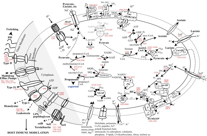
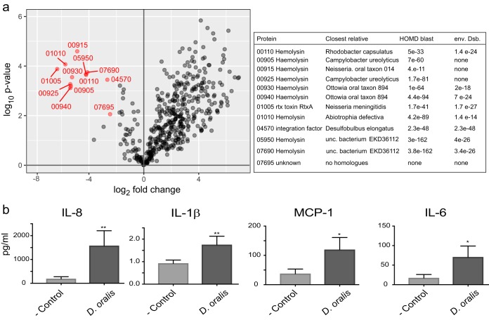
The human oral microbiota encompasses representatives of many bacterial lineages that have not yet been cultured. Here we describe the isolation and characterization of previously uncultured Desulfobulbus oralis, the first human-associated representative of its genus. As mammalian-associated microbes rarely have free-living close relatives, D. oralis provides opportunities to study how bacteria adapt and evolve within a host. This sulfate-reducing deltaproteobacterium has adapted to the human oral subgingival niche by curtailing its physiological repertoire, losing some biosynthetic abilities and metabolic independence, and by dramatically reducing environmental sensing and signaling capabilities. The genes that enable free-living Desulfobulbus to synthesize the potent neurotoxin methylmercury were also lost by D. oralis, a notably positive outcome of host association. However, horizontal gene acquisitions from other members of the microbiota provided novel mechanisms of interaction with the human host, including toxins like leukotoxin and hemolysins. Proteomic and transcriptomic analysis revealed that most of those factors are actively expressed, including in the subgingival environment, and some are secreted. Similar to other known oral pathobionts, D. oralis can trigger a proinflammatory response in oral epithelial cells, suggesting a direct role in the development of periodontal disease.IMPORTANCE Animal-associated microbiota likely assembled as a result of numerous independent colonization events by free-living microbes followed by coevolution with their host and other microbes. Through specific adaptation to various body sites and physiological niches, microbes have a wide range of contributions, from beneficial to disease causing. Desulfobulbus oralis provides insights into genomic and physiological transformations associated with transition from an open environment to a host-dependent lifestyle and the emergence of pathogenicity. Through a multifaceted mechanism triggering a proinflammatory response, D. oralis is a novel periodontal pathobiont. Even though culture-independent approaches can provide insights into the potential role of the human microbiome "dark matter," cultivation and experimental characterization remain important to studying the roles of individual organisms in health and disease.

Keywords: evolution; genome analysis; oral microbiology; periodontitis; proteomics.


Figures

FIG 1
FIG 1
Desulfobulbus sp. strain HOT041 (D. oralis) in coculture with Fusobacterium nucleatum and in pure culture. (A) FISH using fluorescent oligonucleotide probes specific for Deltaproteobacteria (green) and universal Bacteria (red). (B). Growth of Desulfobulbus sp. strain HOT041 and F. nucleatum in coculture monitored by species-specific qPCR (with error bars based on three replicates). (C and D) Scanning electron micrographs of the D. oralis isolate. The arrowheads point to membrane vesicles.
FIG 2
FIG 2
Growth of D. oralis by respiration and by fermentation. (A) Time course cultivation using sulfate and lactate as the electron donor-acceptor pair, with the release of acetate. (B) Substrate utilization and metabolic by-products of pyruvate fermentation. The error bars are based on three culture replicates.
FIG 3
FIG 3
Maximum likelihood phylogeny of Desulfobulbus and related Deltaproteobacteria. The tree is based on full-length SSU rRNA gene sequences. Node support codes are based on 100 bootstrap replicates.
FIG 4
FIG 4
Genomic comparison of D. oralis and D. propionicus. Matching orthologs are connected by internal arc lines, the color scale indicating the degree of pairwise sequence identity at the protein level. External tick lines indicate genes in each genome that contain protein domains enriched (blue) or depleted (red) in D. oralis (based on data in Data Set S2). The outer histograms (light blue) indicate the relative abundance of each detected protein in D. oralis cells (bottom) or culture medium (top). Secreted proteins significantly enriched in the medium are identified and indicated by purple asterisks and lines.
FIG 5
FIG 5
Metabolic reconstruction of D. oralis, with emphasis on membrane processes, energy conservation, and pathogenicity potential. Selected genes encoding enzymes, complexes, and other proteins proposed to function in such cellular processes are indicated by numbers in red, corresponding to GenBank genes in Data Set S1. The putative EtfAB-Ldh complex is depicted in gray.
FIG 6
FIG 6
Proteomics and cytokine stimulation implicating D. oralis in proinflammatory responses. (a) D. oralis proteins that are secreted from the cell (indicated in red) based on enrichment analysis. Only proteins that had at least a 4-fold enrichment and P < 0.01 (based on three biological replicates) were considered. The table on the right identifies those secreted proteins and compares them to their closest homologues in the human oral microbiota or the free-living Desulfobulbus. (b) Cytokine stimulation in oral epithelial keratinocytes by D. oralis cells. Keratinocytes were incubated with D. oralis cells or with the medium control for 48 h, and cytokine stimulation was measured using a multiplex bead assay. Each bar represents the mean ± standard deviation of the mean from triplicate measurements. Asterisks indicate results significantly different from the keratinocyte medium-only control: *, P < 0.05; **, P < 0.01.

References

    1. Dye BA. 2012. Global periodontal disease epidemiology. Periodontol 2000 58:10–25. doi: 10.1111/j.1600-0757.2011.00413.x. - DOI - PubMed
    1. Kassebaum NJ, Bernabé E, Dahiya M, Bhandari B, Murray CJ, Marcenes W. 2014. Global burden of severe periodontitis in 1990–2010: a systematic review and meta-regression. J Dent Res 93:1045–1053. doi: 10.1177/0022034514552491. - DOI - PMC - PubMed
    1. Darveau RP. 2010. Periodontitis: a polymicrobial disruption of host homeostasis. Nat Rev Microbiol 8:481–490. doi: 10.1038/nrmicro2337. - DOI - PubMed
    1. Hajishengallis G, Lamont RJ. 2012. Beyond the red complex and into more complexity: the polymicrobial synergy and dysbiosis (PSD) model of periodontal disease etiology. Mol Oral Microbiol 27:409–419. doi: 10.1111/j.2041-1014.2012.00663.x. - DOI - PMC - PubMed
    1. Hajishengallis G, Liang S, Payne MA, Hashim A, Jotwani R, Eskan MA, McIntosh ML, Alsam A, Kirkwood KL, Lambris JD, Darveau RP, Curtis MA. 2011. Low-abundance biofilm species orchestrates inflammatory periodontal disease through the commensal microbiota and complement. Cell Host Microbe 10:497–506. doi: 10.1016/j.chom.2011.10.006. - DOI - PMC - PubMed

Publication types