. 2018 Mar 13;9(2):e02125-17.
doi: 10.1128/mBio.02125-17.

Human Memory B Cells Targeting Staphylococcus aureus Exotoxins Are Prevalent with Skin and Soft Tissue Infection

Affiliations

Human Memory B Cells Targeting Staphylococcus aureus Exotoxins Are Prevalent with Skin and Soft Tissue Infection

Adam J Pelzek et al. mBio. .

Abstract

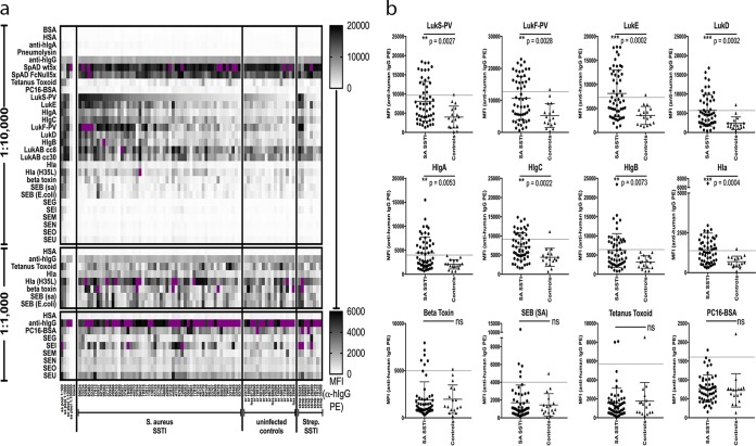
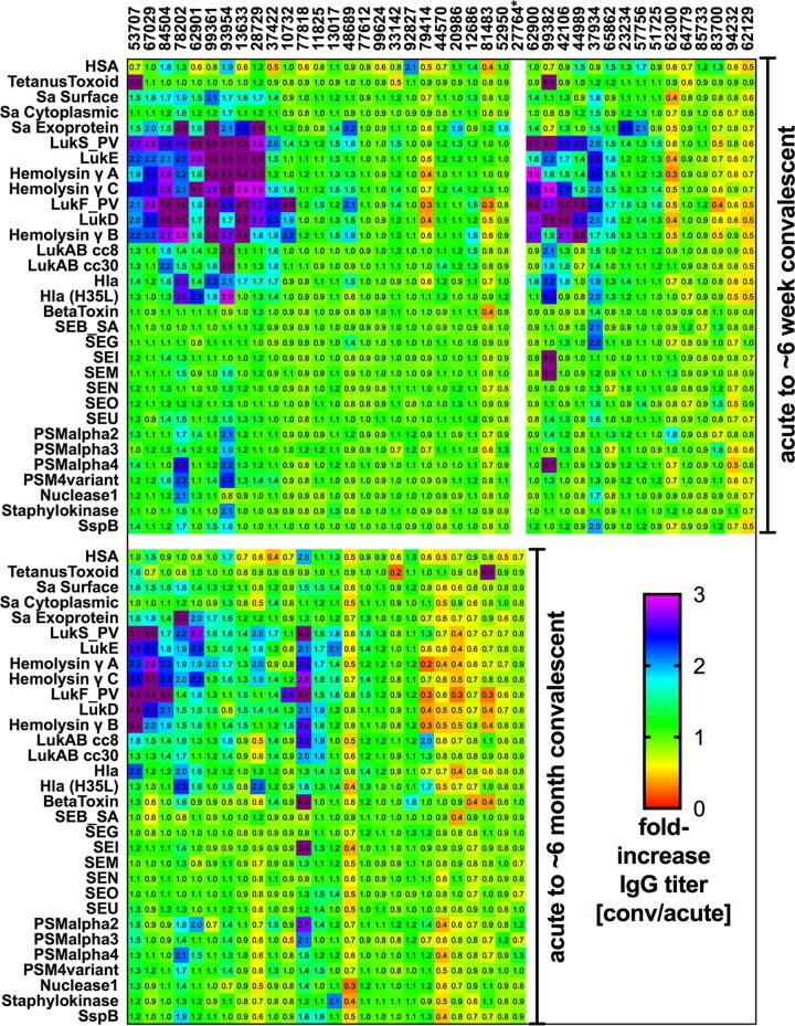
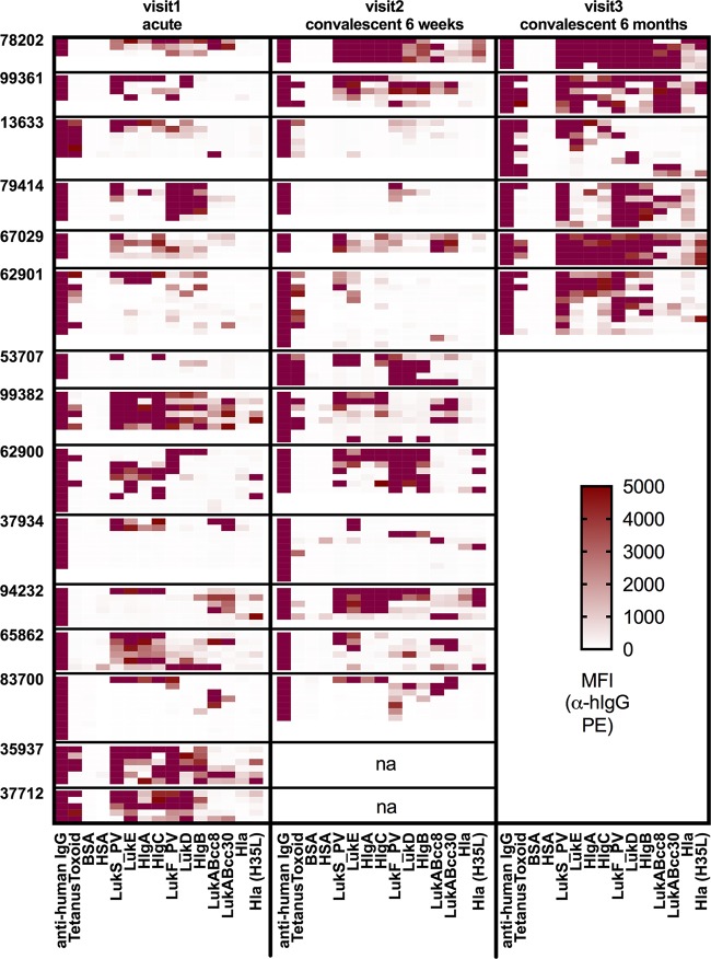
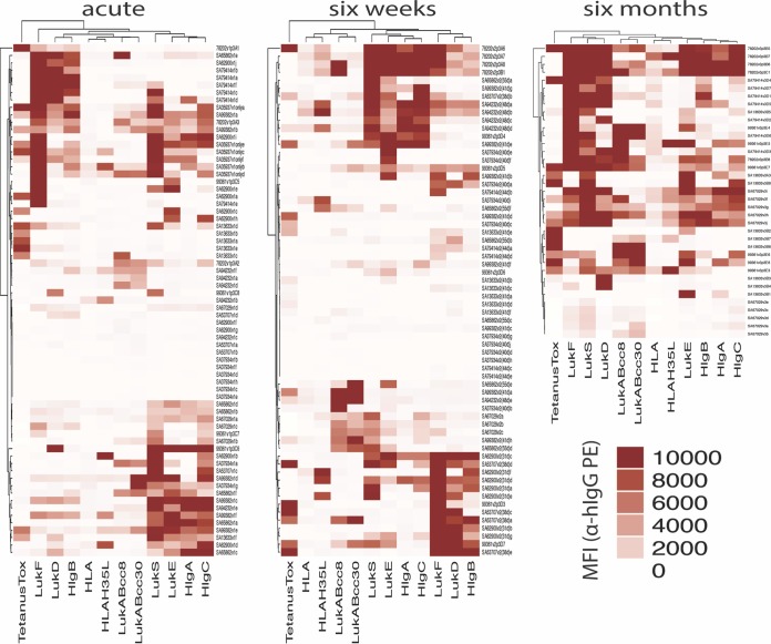
Staphylococcus aureus is a Gram-positive opportunistic pathogen that causes superficial and invasive infections in the hospital and community. High mortality from infection emphasizes the need for improved methods for prevention and treatment. Although S. aureus possesses an arsenal of virulence factors that contribute to evasion of host defenses, few studies have examined long-term humoral and B-cell responses. Adults with acute-phase skin and soft tissue infections were recruited; blood samples were obtained; and S. aureus isolates, including methicillin-resistant strains, were subjected to genomic sequence analysis. In comparisons of acute-phase sera with convalescent-phase sera, a minority (37.5%) of patients displayed 2-fold or greater increases in antibody titers against three or more S. aureus antigens, whereas nearly half exhibited no changes, despite the presence of toxin genes in most infecting strains. Moreover, enhanced antibody responses waned over time, which could reflect a defect in B-cell memory or long-lived plasma cells. However, memory B cells reactive with a range of S. aureus antigens were prevalent at both acute-phase and convalescent-phase time points. While some memory B cells exhibited toxin-specific binding, those cross-reactive with structurally related leucocidin subunits were dominant across patients, suggesting the targeting of conserved epitopes. Memory B-cell reactivity correlated with serum antibody levels for selected S. aureus exotoxins, suggesting a relationship between the cellular and humoral compartments. Overall, although there was no global defect in the representation of anti-S. aureus memory B cells, there was evidence of restrictions in the range of epitopes recognized, which may suggest potential therapeutic approaches for augmenting host defenses.IMPORTANCE The contribution of B-cell memory and long-term antibody responses to host defenses against S. aureus exotoxins remains poorly understood. Our studies confirmed that infection did not commonly lead to enhanced long-term humoral responses. Whereas circulating memory B cells against S. aureus secreted exotoxins were prevalent, they were dominated by cross-reactivity with structurally related leucocidin subunits, consistent with recognition of conserved epitopes. These findings also provide the first evidence of a relationship between the reactivity of antistaphylococcal circulating memory B cells and serum antibody levels. In general, infection was not associated with a global defect in B-cell memory for S. aureus secreted factors, and responses were highly dominated by cross-reactivity to structurally related exotoxins, which arguably may alone be suboptimal in providing host defenses. Our studies illuminate aspects of the S. aureus-host relationship that may better inform strategies for the development of an effective protective vaccine.

Keywords: MRSA; Staphylococcus aureus; antibodies; host response; host-pathogen interactions; leukocidins; memory B cells; pore-forming toxins; skin and soft tissue infection (SSTI); superantigens.


Figures

FIG 1
FIG 1
S. aureus antigen-reactive IgG levels are often higher in infected patients at acute-phase presentation than in healthy controls. Antigen-reactive IgG antibodies in patient serum samples were studied at multiple dilutions by multiplex bead-based antigen array for S. aureus SSTI patients (n = 54), uninfected controls (n = 17), and streptococcal SSTI patients (n = 8). (a) Data are presented as median fluorescence intensity (MFI) values for the 1:10,000 dilution, organized from highest to lowest within each group by reactivity to LukS, with data also shown for the 1:1,000 dilution for the antigen sets with lower IgG reactivity. Purple coloring denotes a signal above the upper limit of the scale. HSA, heat-stabile antigen. (b) Comparisons between S. aureus SSTI patients and uninfected controls are presented for the 1:10,000 serum dilution. The dashed line in each panel represents the mean plus 2 standard deviations for the uninfected control samples; the levels seen with SSTI samples above this threshold were considered to be elevated above control levels. Significance was determined using the Mann-Whitney test (ns, P > 0.05; *, P ≤ 0.05; **, P ≤ 0.01; ***, P ≤ 0.001.
FIG 2
FIG 2
S. aureus skin and soft tissue infection induces longitudinal modulation of IgG levels against pore-forming exotoxin antigens in a subset of individuals. Patient serum samples were studied for IgG at 10-fold dilutions from 1:100 to 1:100,000 by multiplex antigen array. Median fluorescence intensity (MFI) values from all available visits were plotted for each individual for each antigen, and a high-throughput method was developed to assess antigen-reactive titers from the first visit (v1 [acute-phase infection]) to the second visit (v2 [at 6 weeks; n = 40]) and/or the third visit (v3 [at 6 months; n = 26]). Twenty-five subjects completed all visits. Fold changes in titer are presented here for each antigen, representing differences calculated with the following equation: convalescent-phase-sample value/acute-phase-sample value. The cutoff for an increased IgG response was set at a 2-fold increase from the acute-phase baseline, with relative decreases represented as values below 1. *, subject 27764 returned only for the 6-month visit.
FIG 3
FIG 3
IgG-expressing memory B cells reactive with S. aureus exotoxin antigens are abundant in the peripheral blood of S. aureus-infected patients. PBMC (2 × 10⁵ cells/well) from S. aureus SSTI patients (n = 15) were cultured after isolation or following cryopreservation and thaw for 6 days with CpG2006/IL-21/sCD40L stimulation for acute-phase, 6-week convalescent-phase, and 6-month convalescent-phase samples. Supernatants containing antibodies were assessed at a 1:3 dilution for antigen-reactive IgG reactivity by bead-based multiplex assay. Each well is represented as one row, and data are reported as median fluorescence intensity (MFI) values. Purple coloring signifies MFI above the upper limit (>10,000 MFI).
FIG 4
FIG 4
Induced memory B-cell cultures from S. aureus skin and soft tissue infection patients contain IgG that targets structurally homologous leucocidin components. Median fluorescence intensity (MFI) signals for antigen-reactive IgG from induced PBMC cultures from S. aureus SSTI patients at acute-phase and convalescent-phase visits were hierarchically clustered by the Euclidean method in R Studio using the program Complex heatmap. The Pearson clustering method provided similar results (not shown). Culture supernatants from acute-phase samples (visit 1, n = 12 patients), 6-week convalescent-phase samples (visit 2, n = 11), and 6-month convalescent-phase samples (visit 3, n = 5) were included in this analysis.
FIG 5
FIG 5
Cross-reactive antibodies in serum and induced memory B-cell supernatants against “S” or “F” leucocidin components can be inhibited by soluble antigen. A convalescent-phase S. aureus SSTI patient serum pool (n = 20 patients, 6 weeks convalescence) was diluted 1:1,000 and 1:10,000, and 90 µl diluted serum was treated with 10 µl of 1% BSA–PBS buffer (no antigen), LukF, or LukS and then assayed by multiplex bead-based array. For induced PBMC culture supernatants from two S. aureus SSTI patients (79414 and 94232) and one control supernatant with IgG reactive with tetanus toxoid (28531), 40 µl supernatant was added to 50 µl of 1% BSA–PBS and treated as described above. Median fluorescence intensity (MFI) values are shown.

References

    1. Lowy FD. 1998. Staphylococcus aureus infections. N Engl J Med 339:520–532. doi:10.1056/NEJM199808203390806. - DOI - PubMed
    1. Salgado-Pabón W, Schlievert PM. 2014. Models matter: the search for an effective Staphylococcus aureus vaccine. Nat Rev Microbiol 12:585–591. doi:10.1038/nrmicro3308. - DOI - PubMed
    1. Lee BY, Singh A, David MZ, Bartsch SM, Slayton RB, Huang SS, Zimmer SM, Potter MA, Macal CM, Lauderdale DS, Miller LG, Daum RS. 2013. The economic burden of community-associated methicillin-resistant Staphylococcus aureus (CA-MRSA). Clin Microbiol Infect 19:528–536. doi:10.1111/j.1469-0691.2012.03914.x. - DOI - PMC - PubMed
    1. Messina JA, Thaden JT, Sharma-Kuinkel BK, Fowler VG Jr.. 2016. Impact of bacterial and human genetic variation on Staphylococcus aureus infections. PLoS Pathog 12:e1005330. doi:10.1371/journal.ppat.1005330. - DOI - PMC - PubMed
    1. Shopsin B, Kaveri SV, Bayry J. 2016. Tackling difficult Staphylococcus aureus infections: antibodies show the way. Cell Host Microbe 20:555–557. doi:10.1016/j.chom.2016.10.018. - DOI - PubMed

Publication types

MeSH terms

LinkOut - more resources