. 2018 Mar 13;9(1):1046.
doi: 10.1038/s41467-018-03332-7.

Role of monkeys in the sylvatic cycle of chikungunya virus in Senegal

Affiliations

Role of monkeys in the sylvatic cycle of chikungunya virus in Senegal

Benjamin M Althouse et al. Nat Commun. .

Abstract

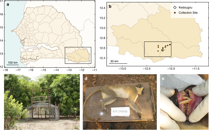
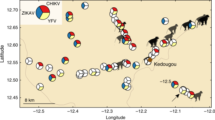
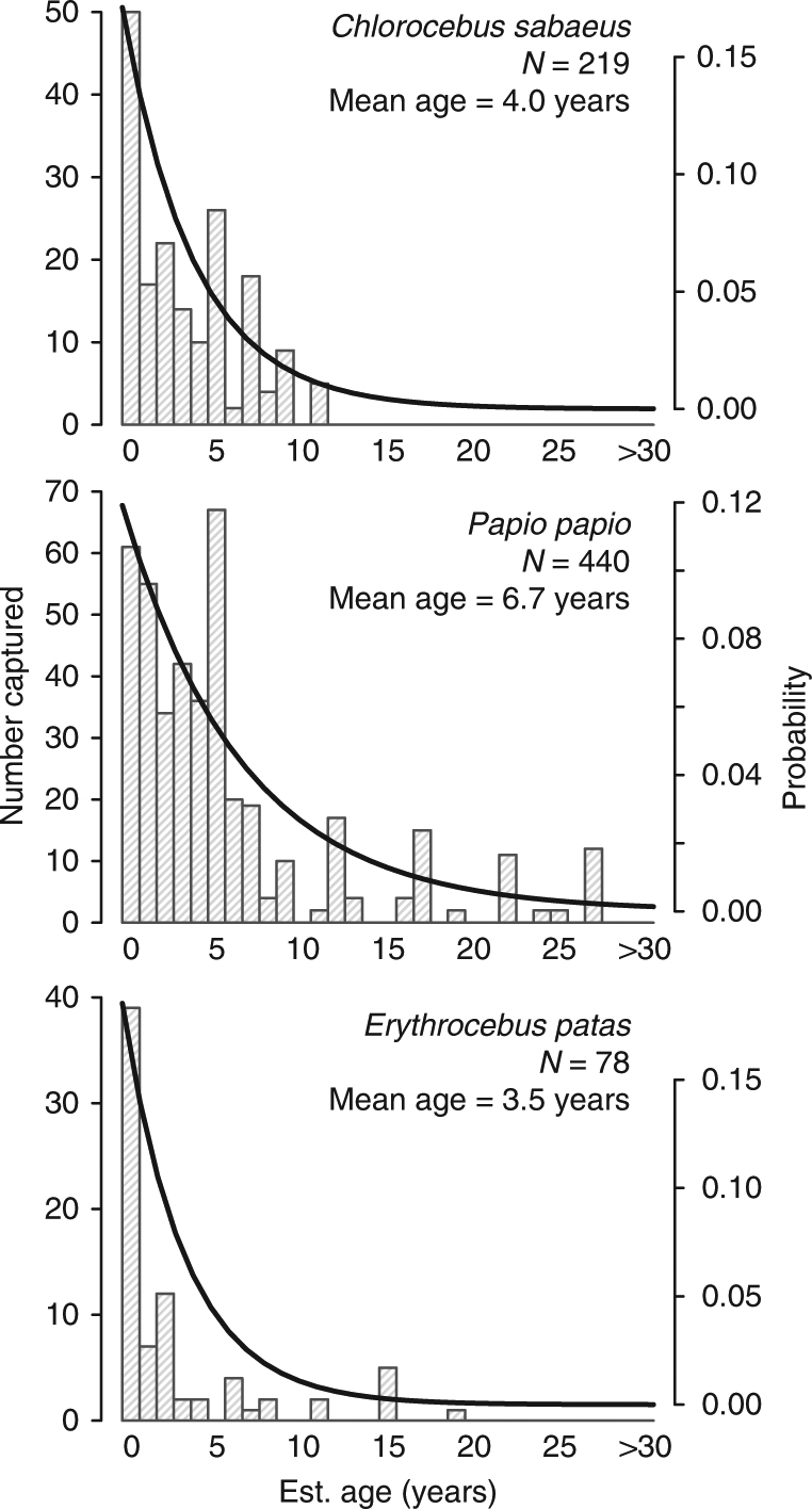
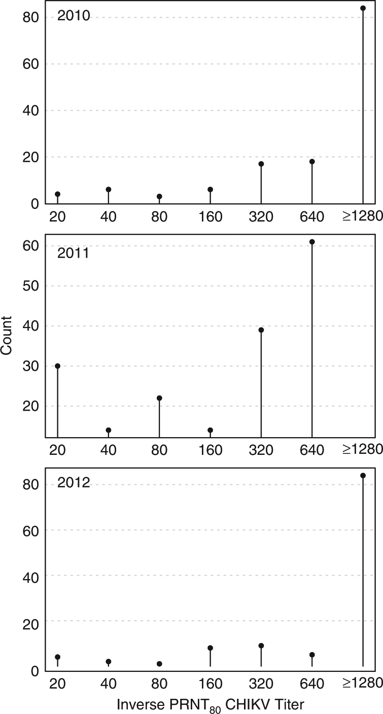
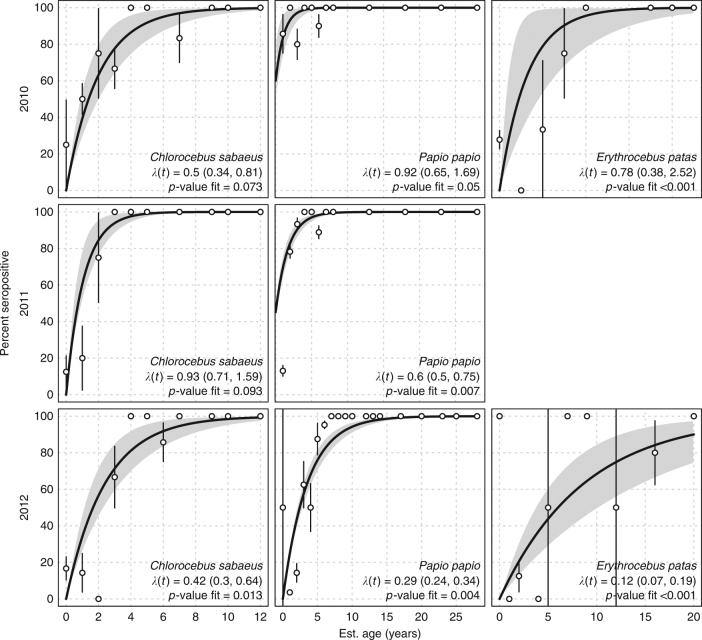
Arboviruses spillover into humans either as a one-step jump from a reservoir host species into humans or as a two-step jump from the reservoir to an amplification host species and thence to humans. Little is known about arbovirus transmission dynamics in reservoir and amplification hosts. Here we elucidate the role of monkeys in the sylvatic, enzootic cycle of chikungunya virus (CHIKV) in the region around Kédougou, Senegal. Over 3 years, 737 monkeys were captured, aged using anthropometry and dentition, and tested for exposure to CHIKV by detection of neutralizing antibodies. Infant monkeys were positive for CHIKV even when the virus was not detected in a concurrent survey of mosquitoes and when population immunity was too high for monkeys alone to support continuous transmission. We conclude that monkeys in this region serve as amplification hosts of CHIKV. Additional efforts are needed to identify other hosts capable of supporting continuous circulation.


Conflict of interest statement

The authors declare no competing interests.

Figures

Fig. 1
Fig. 1
Monkey collection sites and sample individual. a A map of Senegal with the Kédougou Department boxed. b A map of the Kédougou region with the study region boxed and presented in detail in Fig. 2. c A typical trap, and d, e shows a male C. sabaeus estimated to be ~5 years (between 4 and 6 years) of age
Fig. 2
Fig. 2
Distribution of monkey collection sites relative to chikungunya, yellow fever, and zika virus isolations from mosquitoes, 2009–2011. Figure shows the spatial distribution of monkey collection sites (monkey symbols) and the mosquito collection sites (pie charts). Pie slices indicate mosquito collection moving clockwise from 2009 at the top. Red indicates chikungunya virus (CHIKV) mosquito isolates in 2009, yellow indicates yellow fever virus (YFV) mosquito isolates in 2010, and blue indicates Zika virus (ZIKV) mosquito isolates in 2011; unfilled (white) slices indicate that there was no virus isolation in that year. Diamond indicates Kédougou town. Arrow indicates a ZIKV mosquito isolate that is obscured
Fig. 3
Fig. 3
Age distributions of collected monkeys. Panels show the observed age distributions of collected monkeys with exponential distributions (thick line) with rates equal to the mean age of collected individuals, for Chlorocebus sabaeus, Papio papio, and Erythrocebus patas, respectively
Fig. 4
Fig. 4
Inverse plaque reduction neutralization tests (PRNT80) CHIKV titers by year. Figure shows number of animals (counts) per antibody titer by year for all three species of monkey
Fig. 5
Fig. 5
Forces of infection by species and year. Panels show the forces infection (λ(t)) and p-values (Chi-squared test comparing model to saturated model) for the fit across years for C. sabaeus, E. patas, and P. papio, respectively. Too few E. patas were collected in 2011 to obtain estimates. We included all monkeys <1-year-old in the 0-age category (we present seropositivity results for monkeys under 3 years of age in Supplementary Fig. 4). Points are the proportion of seropositve monkeys per age year with confidence intervals. Thick black line is the fit of the force of infection, gray bands are bootstrap confidence intervals for the fit

References

    1. Taylor LH, Latham SM, Mark EJ. Risk factors for human disease emergence. Philos. Trans. R. Soc. Lond. B: Biol. Sci. 2001;356:983–989. doi: 10.1098/rstb.2001.0888. - DOI - PMC - PubMed
    1. Vorou R. Zika virus, vectors, reservoirs, amplifying hosts, and their potential to spread worldwide: what we know and what we should investigate urgently. Int. J. Infect. Dis. 2016;48:85–90. doi: 10.1016/j.ijid.2016.05.014. - DOI - PubMed
    1. Weaver, S. C. in Infectious diseases from nature: mechanisms of viral emergence and persistence (eds Peters, C.J. & Calisher, C.H.) 33–44 (Springer, Vienna, 2005).
    1. Reisen WK. Ecology of west nile virus in North America. Viruses. 2013;5:2079–2105. doi: 10.3390/v5092079. - DOI - PMC - PubMed
    1. Rosen L. The natural history of Japanese encephalitis virus. Annu. Rev. Microbiol. 1986;40:395–414. doi: 10.1146/annurev.mi.40.100186.002143. - DOI - PubMed