. 2018 Mar 13;8(1):4407.
doi: 10.1038/s41598-018-22729-4.

Direct coupling of detergent purified human mGlu5 receptor to the heterotrimeric G proteins Gq and Gs

Affiliations

Direct coupling of detergent purified human mGlu5 receptor to the heterotrimeric G proteins Gq and Gs

Chady Nasrallah et al. Sci Rep. .

Abstract

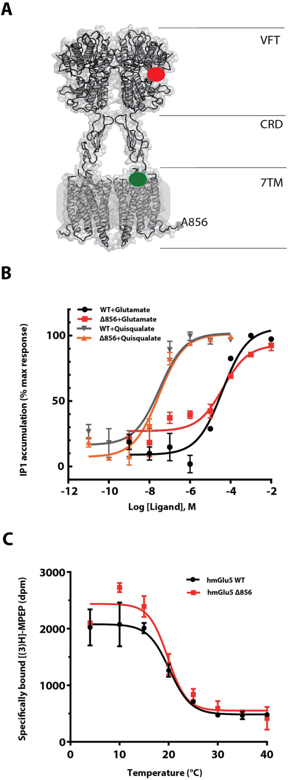
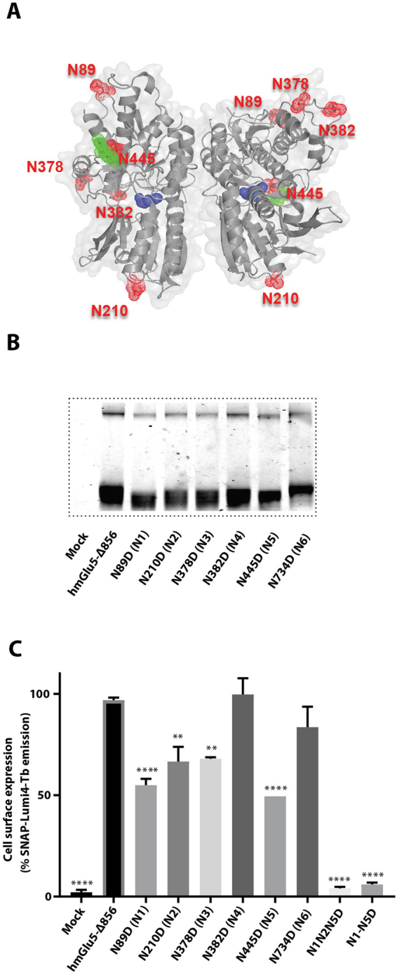
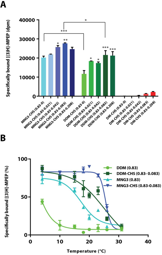
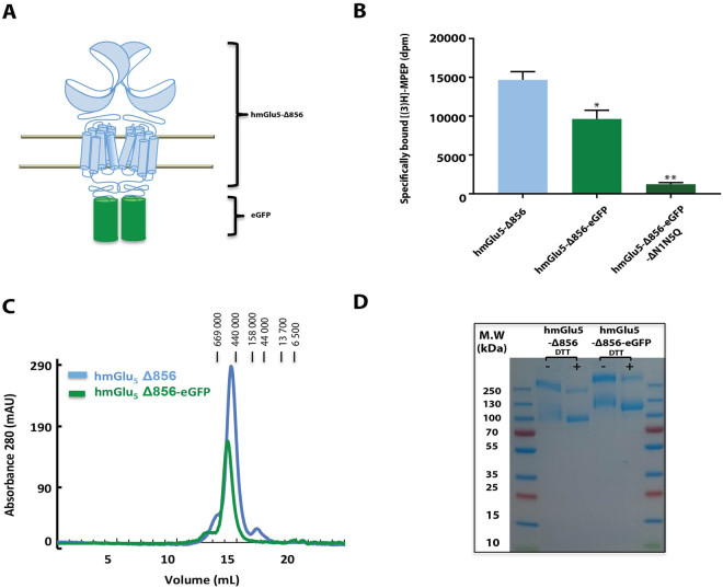
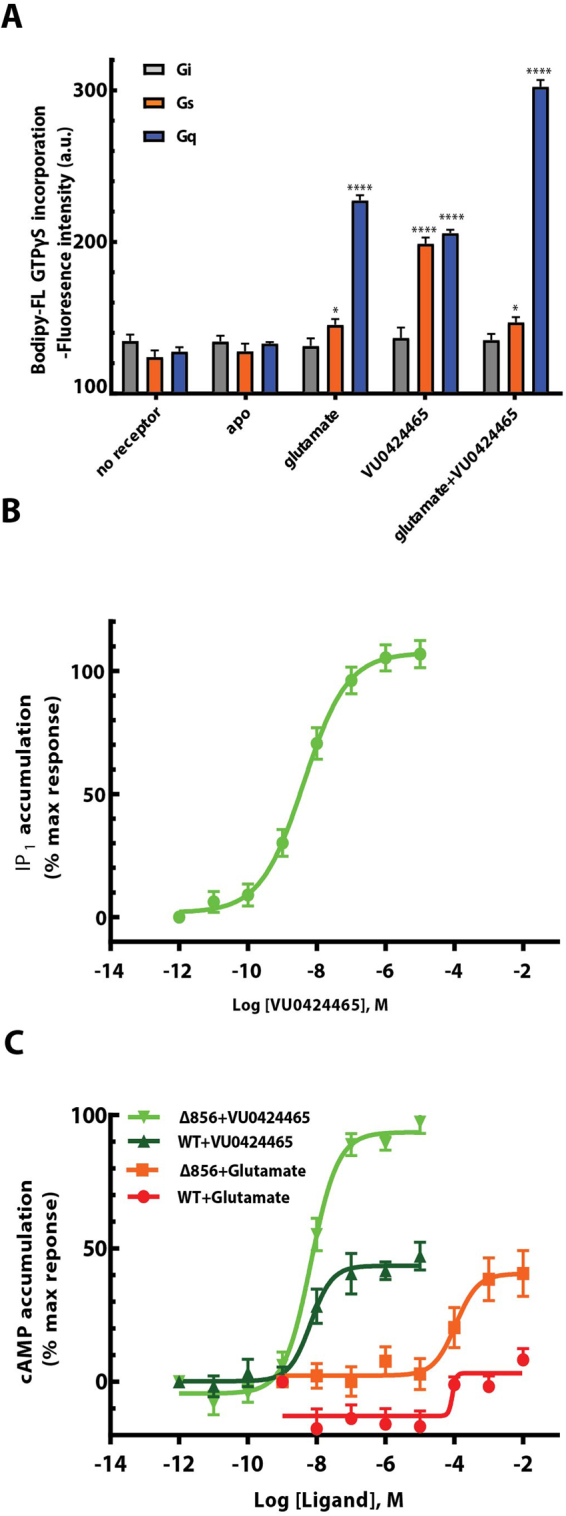
The metabotropic glutamate (mGlu) receptors are class C G protein-coupled receptors (GPCRs) that modulate synaptic activity and plasticity throughout the mammalian brain. Signal transduction is initiated by glutamate binding to the venus flytrap domains (VFT), which initiates a conformational change that is transmitted to the conserved heptahelical domains (7TM) and results ultimately in the activation of intracellular G proteins. While both mGlu1 and mGlu5 activate Gαq G-proteins, they also increase intracellular cAMP concentration through an unknown mechanism. To study directly the G protein coupling properties of the human mGlu5 receptor homodimer, we purified the full-length receptor, which required careful optimisation of the expression, N-glycosylation and purification. We successfully purified functional mGlu5 that activated the heterotrimeric G protein Gq. The high-affinity agonist-PAM VU0424465 also activated the purified receptor in the absence of an orthosteric agonist. In addition, it was found that purified mGlu5 was capable of activating the G protein Gs either upon stimulation with VU0424465 or glutamate, although the later induced a much weaker response. Our findings provide important mechanistic insights into mGlu5 G protein-dependent activity and selectivity.


Conflict of interest statement

The authors declare no competing interests.

Figures

Figure 1
Figure 1
Functional C-terminally truncated SNAP-mGlu5 receptor dimer. (A) 3-Dimensional model of the human mGlu5 receptor truncated after residue Ala856. The truncated receptor is composed of all segments required for effective G protein signaling, including the Venus Flytrap domain (VFT), Cysteine-Rich Domain (CRD) and the heptahelical transmembrane domain (7TM). The red and green spheres represent the orthosteric and allosteric binding sites, respectively. (B) Glutamate- and quisqualate-induced IP1 production in HEK293 cells transiently transfected with SNAP-Tag full-length human mGlu5 receptor (black and grey curves) or SNAP-Tag C-terminal mGlu5-Δ856 receptor (red and orange curves), respectively. (C) Thermal stability of the full-length wild-type mGlu5 and mGlu5-Δ856 [3H]-MPEP-bound receptors. Data points for both assays represent the mean ± SEM of three independent experiments.
Figure 2
Figure 2
Characterization of the SNAP-mGlu5-∆856 N-glycosylation profile in HEK293 cells. (A) The five mGlu5 N-linked glycosylation sites are highlighted as surface spheres (red) and the sugar moieties in green on the cartoon structure (grey) of the VFT (code pdb 3LMK). Atoms in the glutamate molecules bound to the VFT are represented as blue spheres. (B) SDS-PAGE gel migration of monomeric SNAP-mGlu5-Δ856 and different N-glycosylation mutants. Samples were treated with reducing agent (DTT). (C) Cell surface expression of the fluorescently labeled SNAP-mGlu5-Δ856 and different N-glycosylation mutants using SNAP-Lumi4Tb. Gel is representative of one experiment repeated at least two time. Expression profiles are representative of at least three separate experiments. Dunnett’s test as part of one-way ANOVA was used for comparison with hmGlu5-Δ856 set as reference level.
Figure 3
Figure 3
Detergent solubilisation and thermal stability of mGlu5-Δ856 [3H]-MPEP-bound receptor in insect cells. (A) Quantification of unpurified mGlu5-Δ856 receptor solubilized at 4 °C using [3H]-MPEP binding assay measured in a series of maltoside-based detergents including lauryl maltose neopentyl glycol-3 (MNG3), n-Dodecyl-β-D-Maltoside (DDM) and n-Decyl-β-D-Maltoside (DM) supplemented with different ratio of cholesteryl hemisuccinate (CHS). (B) Thermal stability of the mGlu5-Δ856 [3H]-MPEP-bound receptor solubilized in four different conditions; DDM (0.83%), DDM-CHS (0.83–0.083%), MNG3 (0.83%) and MNG3-CHS (0.83–0.083%). Data points for both assays (A,B) represent the mean ± SEM of three independent experiments. Dunnett’s test as part of one-way ANOVA was used for comparison with MNG3-CHS (0.83–0) and DDM-CHS (0.83–0) set as reference level for MNG3 and DDM supplemented with CHS, respectively.
Figure 4
Figure 4
Purification of the dimer mGlu5 -∆856 produced in SF9 cells. (A) Cartoon representation of the dimer human mGlu5 receptor (light blue) with the fusion protein eGFP (green) replacing the c-terminal domain at position 856. (B) Quantification of unpurified mGlu5 receptor using [3H]-MPEP binding assay, measured for mGlu5-Δ856, mGlu5-Δ856-eGFP and mGlu5-Δ856-eGFP (N1-N5). Dunnett’s test as part of one-way ANOVA was used for comparison with mGlu5-Δ856 set as reference level. (C) Size exclusion chromatography profile for both constructs, mGlu5-Δ856 (Ve = 14.5 mL) and mGlu5-Δ856-eGFP (Ve = 14.2 mL) using superpose 6 increase. (D) Coomassie-blue stained SDS-PAGE gel of mGlu5-Δ856 and mGlu5-Δ856-eGFP. The gel shown is representative of three independent experiments.
Figure 5
Figure 5
Glutamate and VU0424465 trigger mGlu5-dependent Gs and Gq activation. (A) GTPγS fluorescence binding experiments were carried out within detergent purified receptor induced by ligand activation. The fluorescence baseline is estimated based on the receptor alone without any ligands. Data points represent average of three independent measurements with SEM. Dunnett’s test as part of one-way ANOVA was used for comparison with the apo condition set as reference level. (B) mGlu5-dependent Gq activation was measured using the IP one cell-based assay kit (Cisbio) in HEK293 cells and in the presence of VU0424465 with a pEC50 of 8.53 ± 0.13 (n = 4). (C) mGlu5-dependent Gs activation was measured using the cAMP cell-based assay kit (Cisbio) in HEK293 cells and in the presence of glutamate and VU0424465, independently. The dose response curves show a positive response for VU0424465 at the truncated mGlu5-Δ856 (light green, pEC50 of 8.17 ± 0.11; n = 4) and WT human mGlu5 (Dark green, pEC50 of 7.80 ± 0.50; n = 3) curve. A weaker activation is observed for glutamate at the truncated mGlu5-Δ856 (orange, pEC50 of 3.86 ± 0.20; n = 3) and WT human mGlu5 (red, pEC50 of 3.36 ± 0.72; n = 3) curve. Data points represent the average of triplicate measurements from at least three independent experiments with SEM.

References

    1. Conn PJ, Christopoulos A, Lindsley CW. Allosteric modulators of GPCRs: a novel approach for the treatment of CNS disorders. Nat Rev Drug Discov. 2009;8:41–54. doi: 10.1038/nrd2760. - DOI - PMC - PubMed
    1. Recasens M, Guiramand J, Aimar R, Abdulkarim A, Barbanel G. Metabotropic glutamate receptors as drug targets. Curr Drug Targets. 2007;8:651–681. doi: 10.2174/138945007780618544. - DOI - PubMed
    1. Kniazeff J, Prezeau L, Rondard P, Pin JP, Goudet C. Dimers and beyond: The functional puzzles of class C GPCRs. Pharmacol Ther. 2011;130:9–25. doi: 10.1016/j.pharmthera.2011.01.006. - DOI - PubMed
    1. Urwyler S. Allosteric modulation of family C G-protein-coupled receptors: from molecular insights to therapeutic perspectives. Pharmacol Rev. 2011;63:59–126. doi: 10.1124/pr.109.002501. - DOI - PubMed
    1. Kniazeff J, et al. Closed state of both binding domains of homodimeric mGlu receptors is required for full activity. Nat Struct Mol Biol. 2004;11:706–713. doi: 10.1038/nsmb794. - DOI - PubMed

Publication types

MeSH terms

Substances

LinkOut - more resources