. 2018 Mar 13;8(1):4393.
doi: 10.1038/s41598-018-22828-2.

Fluorescent polydopamine nanoparticles as a probe for zebrafish sensory hair cells targeted in vivo imaging

Affiliations

Fluorescent polydopamine nanoparticles as a probe for zebrafish sensory hair cells targeted in vivo imaging

Gyo Eun Gu et al. Sci Rep. .

Abstract

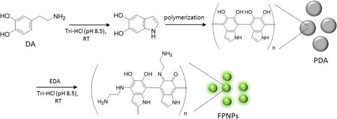
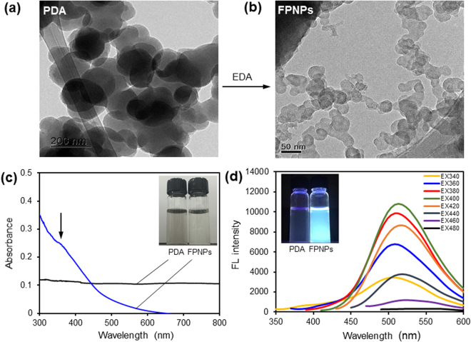
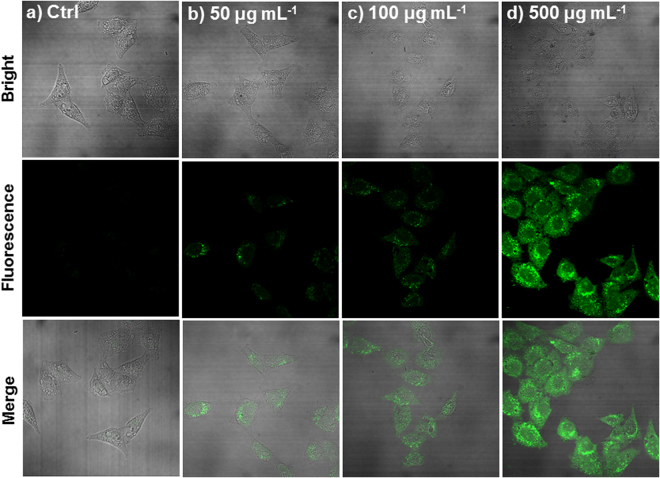
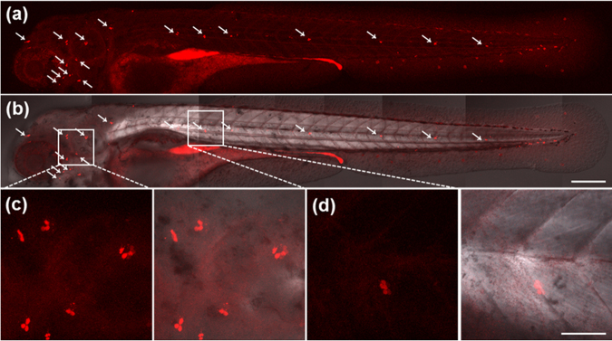
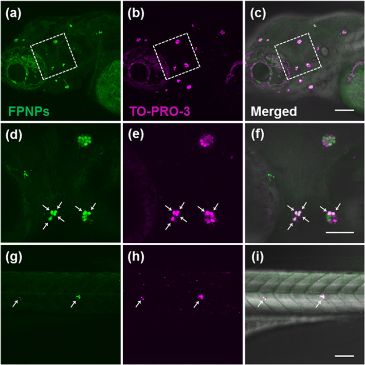
Fluorescent polydopamine nanoparticles (FPNPs) are prepared via the ethylenediamine (EDA)-induced degradation of as-prepared non-fluorescent polydopamine (PDA) and used for targeted bioimaging. The reductive treatment of PDA in the presence of EDA yields fluorescent precipitates, inspiring us to seek various biological approaches to preparing FPNPs with excellent optical and biocompatible properties. Moreover, we firstly found that FPNPs selectively label neuromast hair cells in the lateral line of zebrafish, their applications as a reliable fluorescent indicator to investigate the neuromast hair cells, to in turn determine the viability of hair cells, was demonstrated. FPNPs also provided a minimal toxicity enable to assay the number of functional hair cells per neuromast in live animals as development proceeds. Upon combined incubation with TO-PRO-3, a well-established hair cell marker, all hair cells that were rapidly labeled with FPNPs were observed to be also completely labeled with the TO-PRO-3, labeling hair cells in neuromasts positioned in the supraorbital, otic and occipital lateral line as well as in posterior lateral line of living zebrafish larvae. Their potential efficacy for biological applications was demonstrated by their excellent optical and biocompatible properties, offering new opportunities in cancer research, real-time monitoring of stem cell transplantation and other cell-based therapies.


Conflict of interest statement

The authors declare no competing interests.

Figures

Figure 1
Figure 1
Schematic illustration of the fluorescent polydopamine nanoparticle (FPNP)s preparation process.
Figure 2
Figure 2
(a and b) Transmission electron microscopy (TEM) images of polydopamine (PDA particles; 1a) and fluorescent polydopamine nanoparticles (FPNPs; 1b). (c) Ultraviolet-visible spectroscopy (UV-vis) absorption (black) and FPNPs (blue) in water and (d) FL emission spectra with gradually increased excitation wavelengths from 340 nm to 480 nm after dispersion of the FPNPs in water (50 μg mL−1).
Figure 3
Figure 3
Confocal fluorescence images of living HeLa cells incubated with FPNPs of 0 μg mL−1, (b) 50 μg mL−1, (c) 100 μg mL−1 and (d) 500 μg mL−1. Incubation was performed at 37 °C under a humidified atmosphere containing 5% CO2. The fluorescence was determined at 488 nm with excitation at 405 nm.
Figure 4
Figure 4
Sensory hair cells in the lateral line of whole zebrafish larvae at 4 dpf. White arrows (a and b) show the lateral line hair cells in a zebrafish larva incubated with FPNPs (2 mg mL−1) from 2 to 4 dpf. (a) Fluorescence image, (b) Merged image with DIC. Enlarged images of (c) cranial neuromasts and (d) trunk neuromasts. The fluorescence was determined at 543 nm with excitation at 405 nm. Scale bars: 200 μm (a,b), 50 μm (c,d).
Figure 5
Figure 5
Live hair cells are double labelled with FPNPs (2 mg mL−1) and TO-PRO-3 (2 μM) in zebrafish. The panels (a,d and g) show the cells stained with FPNPs (excitation wavelength at 405 nm, emission wavelength at 488 nm, respectively). The panels (b,e and h) show the fluorescence images by TO-PRO-3 (excitation wavelength at 635 nm, emission wavelength at 647 nm, respectively). The panels (c,f and i) show the overlay images of (a) + (b), (d) + (e), and (g) + (h), respectively. White arrows (d,e, f,g,h and i) indicate the co-stained hair cells. The enlarged images of the white boxed areas in (a,b and c) correspond to (d),(e), and (f), respectively. Scale bars: 50 μm (af), 100 μm (ac and gi).

References

    1. Li K, et al. Photostable fluorescent organic dots with aggregation-induced emission (AIE dots) for noninvasive long-term cell tracing. Sci. Rep. 2013;3:1150. doi: 10.1038/srep01150. - DOI - PMC - PubMed
    1. Xu X, Liu R, Li L. Nanoparticles made of π-conjugated compounds targeted for chemical and biological applications. Chem. Commun. 2015;51:16733–16749. doi: 10.1039/C5CC06439B. - DOI - PubMed
    1. Schill J, Schenning APHJ, Brunsveld L. Self-Assembled Fluorescent Nanoparticles from π-Conjugated Small Molecules: En Route to Biological Applications. Macromol. Rapid Commun. 2015;36:1306–1321. doi: 10.1002/marc.201500117. - DOI - PubMed
    1. Kaeser A, Schenning APHJ. Fluorescent Nanoparticles Based on Self-Assembled π-Conjugated Systems. Adv. Mater. 2010;22:2985–2997. doi: 10.1002/adma.201000427. - DOI - PubMed
    1. Zhang X, et al. Aggregation induced emission-based fluorescent nanoparticles: fabrication methodologies and biomedical applications. J. Mater. Chem. B. 2014;2:4398–4414. doi: 10.1039/c4tb00291a. - DOI - PubMed

Publication types