Review
. 2018 Jul;32(7):1500-1514.
doi: 10.1038/s41375-018-0061-9. Epub 2018 Feb 22.

Mesenchymal stem cells in multiple myeloma: a therapeutical tool or target?

Affiliations
Review

Mesenchymal stem cells in multiple myeloma: a therapeutical tool or target?

Song Xu et al. Leukemia. 2018 Jul.

Abstract

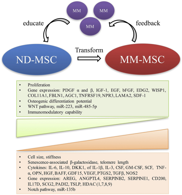
Multiple myeloma (MM) is a malignant plasma cell (PC) disorder, characterized by a complex interactive network of tumour cells and the bone marrow (BM) stromal microenvironment, contributing to MM cell survival, proliferation and chemoresistance. Mesenchymal stem cells (MSCs) represent the predominant stem cell population of the bone marrow stroma, capable of differentiating into multiple cell lineages, including fibroblasts, adipocytes, chondrocytes and osteoblasts. MSCs can migrate towards primary tumours and metastatic sites, implying that these cells might modulate tumour growth and metastasis. However, this issue remains controversial and is not well understood. Interestingly, several recent studies have shown functional abnormalities of MM patient-derived MSCs indicating that MSCs are not just by-standers in the BM microenvironment but rather active players in the pathophysiology of this disease. It appears that the complex interaction of MSCs and MM cells is critical for MM development and disease outcome. This review will focus on the current understanding of the biological role of MSCs in MM as well as the potential utility of MSC-based therapies in this malignancy.


Conflict of interest statement

The authors declare that they have no conflict of interest.

Figures

Fig. 1
Fig. 1
Biological alterations of MSCs in the MM tumour microenvironment. It can be assumed that normal MSCs are educated by MM cells and transform into MM-MSCs, which in turn influence MM cell growth. It cannot be excluded that some abnormalities are intrinsic (and not MM cell-induced). MSC mesenchymal stem cell, MM multiple myeloma, PDGF platelet-derived growth factor, IGF1 insulin-like growth factor 1, EGF epidermal growth factor, bFGF basic fibroblast growth factor, EDG2 endothelial differentiation, lysophosphatidic acid G-protein-coupled receptor 2, WISP1 WNT1-induced secreted protein-1, COL11A1 collagen type XI α1 chain, SDF1 stromal derived factor-1, FBLN1 fibulin 1, AGC1 amino acid transporter AGC1, TNFRSF19 TNF receptor superfamily member 19, NPR3 natriuretic peptide receptor 3, LAMA2 laminin subunit α2, IL interleukin, DKK1 dickkopf-1, CSF colony stimulating factor, SCF stem cell factor, TNF-α tumour necrosis factor-α, OPN osteopontin, HGF hepatocyte growth factor, VEGF vascular endothelial growth factor, BAFF B cell-activating factor, GDF15 growth differentiation factor 15, PTGS2 prostaglandin-endoperoxide synthase 2, TGFβ transforming growth factor-β, NOS2 nitric oxide synthase 2, AREG amphiregulin, ANGPTL4 angiopoietin like 4, SERPINB2 serpin family B member 2, SERPINE1 serpin family E member 1, SCG2 secretogranin II, PADI2 peptidyl arginine deiminase 2, TSLP thymic stromal lymphopoietin, HDAC histone deacetylase
Fig. 2
Fig. 2
Schematic diagram of MSC interactions in MM tumour microenvironment. Direct and indirect interactions with MM cells induce MSCs to acquire abnormal phenotypes, which in turn lead to the formation of BM microenvironment influencing MM tumour development and progression of osteolytic bone lesions. MSC mesenchymal stem cell, MM multiple myeloma, HGF hepatocyte growth factor, VEGF vascular endothelial growth factor, TGF-β transforming growth factor-β, bFGF basic fibroblast growth factor, IL interleukin, DKK1 Dickkopf-1, Cx43 Connexin-43, SDF1 stromal-derived factor-1, IGF1 insulin-like growth factor 1, TNF-α tumour necrosis factor-α, sFRP secreted frizzled-related protein, CCL3 chemokine (C-C motif) ligand 3, RANKL nuclear factor-κB ligand, DcR3 soluble decoy receptor 3, MMP-13 matrix metalloproteinases 13, OPG osteoprotegerin, Runx2 runt-related transcription factor 2

References

    1. Arnulf B, Lecourt S, Soulier J, Ternaux B, Lacassagne MN, Crinquette A, et al. Phenotypic and functional characterization of bone marrow mesenchymal stem cells derived from patients with multiple myeloma. Leukemia. 2007;21:158–63. - PubMed
    1. Corre J, Mahtouk K, Attal M, Gadelorge M, Huynh A, Fleury-Cappellesso S, et al. Bone marrow mesenchymal stem cells are abnormal in multiple myeloma. Leukemia. 2007;21:1079–88. - PMC - PubMed
    1. Garderet L, Mazurier C, Chapel A, Ernou I, Boutin L, Holy X, et al. Mesenchymal stem cell abnormalities in patients with multiple myeloma. Leuk Lymphoma. 2007;48:2032–41. - PubMed
    1. Wallace SR, Oken MM, Lunetta KL, Panoskaltsis-Mortari A, Masellis AM. Abnormalities of bone marrow mesenchymal cells in multiple myeloma patients. Cancer. 2001;91:1219–30. - PubMed
    1. Zdzisinska B, Bojarska-Junak A, Dmoszynska A, Kandefer-Szerszen M. Abnormal cytokine production by bone marrow stromal cells of multiple myeloma patients in response to RPMI8226 myeloma cells. Arch Immunol Ther Exp (Warsz) 2008;56:207–21. - PMC - PubMed

Publication types

MeSH terms