. 2018 Jan 14:2018:4283672.
doi: 10.1155/2018/4283672. eCollection 2018.

Increase of Vascular Endothelial Growth Factor and Decrease of MCP-1 and Some Updated Epidemiology Aspects of Cystic Echinococcosis Human Cases in Calabria Region

Affiliations

Increase of Vascular Endothelial Growth Factor and Decrease of MCP-1 and Some Updated Epidemiology Aspects of Cystic Echinococcosis Human Cases in Calabria Region

Giovanni Matera et al. Mediators Inflamm. .

Abstract

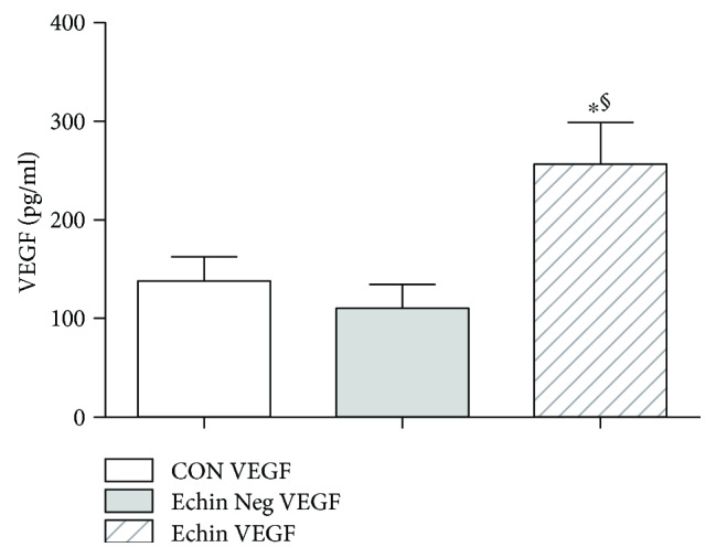
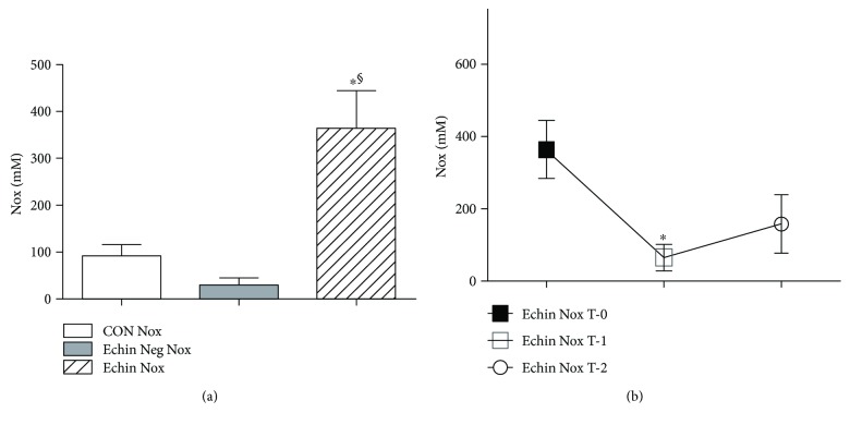
We aim to investigate some of the pathogenetic mediators of the human echinococcosis and to obtain updated epidemiological findings on cases of echinococcosis in Calabria, Southern Italy. Echinococcosis diagnosis was based on imaging, serological investigations, and molecular assay. Indeed, real-time PCR indicated the presence of G2/G3 genotypes of Echinococcus granulosus complex. Regarding pathogenesis, a relevant novel tool of immune depression should be deemed the reduced level of serum MCP-1. Also, we found a previously unreported VEGF, possibly associated with neovascularization requested by the parasite cyst metabolism. Cytokine profiles suggest a bias of the immunity toward Th2 and Treg responses. Nitric oxide levels exhibited a significant decrease one week after therapy versus basal level measured before surgery and/or chemotherapy. An increase of serum total IgE class and IgG4 subclass was found in Echinococcus-positive patients versus controls. Our data demonstrated an endemic spreading, at least in the province of Catanzaro and neighboring Calabria territories, for such parasitosis with the novel issue of the number of female overcoming male cases. In conclusion, the novel findings of this study were the increased VEGF and the reduced serum MCP-1 in the studied cases, as well as the number of Echinococcus-infected females overcoming the infected males.


Figures

Figure 1
Figure 1
(a) PCR real-time curves obtained from cystic liquid of our patient C.A. following the procedure of Capuano et al. [12] reported in “Material and Methods”; (b) melting curves of hydatid cyst of standard E. granulosus genotypes G1 with Tm 76.55 (A), genotypes G2/G3 with Tm 77.03 (B), and of hydatid cyst of patient C.A. with Tm 77.11 (C) from this study. Neg is a negative control.
Figure 2
Figure 2
Serum levels of IL-6 in the group of infected patients (Echin IL-6) are significantly increased (p < 0.05) in comparison to the IL-6 concentration evaluated in the group of Echinococcus-negative patients (Echin Neg IL-6) and the group of control subjects (CON IL-6). p < 0.05 versus CON; §p < 0.05 versus Echin Neg, after ANOVA and Fisher PLSD test.
Figure 3
Figure 3
Serum levels of IL-10 in the group of infected patients (Echin IL-10) are significantly increased (p < 0.05) in comparison to the IL-10 concentration evaluated in the group of Echinococcus-negative patients (Echin Neg IL-10) and in the group of control subjects (CON IL-10). p < 0.05 versus CON, after ANOVA and Fisher PLSD test.
Figure 4
Figure 4
Serum levels in the group of infected patients (Echin MCP-1) are significantly decreased (p < 0.05) versus the correspondent parameters in the group of Echinococcus-negative patients (Echin Neg MCP-1) and in the group of control subjects (CON MCP-1). p < 0.05 versus CON, after ANOVA and Fisher PLSD test.
Figure 5
Figure 5
Serum levels of VEGF in the group of infected patients (Echin VEGF) are significantly increased (p < 0.05) versus the correspondent parameters in the group of Echinococcus-negative patients (Echin Neg VEGF) and in the group of control subjects (CON VEGF). Values are expressed as mean ± SEM. p < 0.05 versus CON; §p < 0.05 versus Echin Neg, after ANOVA and Fisher PLSD test.
Figure 6
Figure 6
(a) Serum levels of Nox showed a significant increase (p < 0.05) compared to corresponding serum concentrations found in healthy controls and Echinococcus-negative patients. Values are expressed as mean ± SEM. p < 0.05 versus CON; §p < 0.05 versus Echin Neg, after ANOVA and Fisher PLSD test. (b) Significant decline in the concentrations of nitric oxide metabolites in parasite-infected patients one week after surgery (time T-1) (p < 0.05) compared with the levels found in patients before any therapy (time T-0); however, 1-2 months after therapy (time T-2), such nitric oxide metabolites exhibited a light increase.
Figure 7
Figure 7
(a) Serum levels of IgG4 in the group of infected patients (Echin IgG4) are significantly increased (p < 0.05) versus the same parameters in the group of Echin-negative patients (Echin Neg IgG4) and in the group of control subjects (CON IgG4). Values are expressed as mean ± SEM. p < 0.05 versus CON; §p < 0.05 versus Echin Neg, after ANOVA and Fisher PLSD test. (b) IgG4 levels showed a significant decrease in parasite-infected patients one week after surgery (time T-1) (p < 0.05) compared with the levels found in patients before any therapy (time T-0); such reduction was observed even 1-2 months after therapy (time T-2). p < 0.05 versus Echin IgG4 T-0, after ANOVA and Fisher PLSD test.
Figure 8
Figure 8
(a) Serum levels of IgE in the group of infected patients showed a significant increase (p < 0.05) compared to corresponding serum concentrations found in healthy controls and Echinococcus-negative patients. Values are expressed as mean ± SEM p < 0.05 versus CON; §p < 0.05 versus Echin Neg, after ANOVA and Fisher PLSD test. (b) A significant decline in the concentrations of levels of IgE in parasite-infected patients one week after surgery (time T-1) (p < 0.05) compared with the levels found in patients before any therapy (time T-0). p < 0.05 versus Echin IgE T-0, after ANOVA and Fisher PLSD test.

References

    1. Thompson R. C. A., Jenkins D. J. Echinococcus as a model system: biology and epidemiology. International Journal for Parasitology. 2014;44(12):865–877. doi: 10.1016/j.ijpara.2014.07.005. - DOI - PubMed
    1. Petrone L., Vanini V., Petruccioli E., et al. IL-4 specific-response in whole blood associates with human cystic echinococcosis and cyst activity. The Journal of Infection. 2015;70(3):299–306. doi: 10.1016/j.jinf.2014.10.009. - DOI - PubMed
    1. Cucher M. A., Macchiaroli N., Baldi G., et al. Cystic echinococcosis in South America: systematic review of species and genotypes of Echinococcus granulosus sensu lato in humans and natural domestic hosts. Tropical Medicine & International Health. 2016;21(2):166–175. doi: 10.1111/tmi.12647. - DOI - PubMed
    1. Brunetti E., Kern P., Vuitton D. A., Writing Panel for the WHO-IWGE Expert consensus for the diagnosis and treatment of cystic and alveolar echinococcosis in humans. Acta Tropica. 2010;114(1):1–16. doi: 10.1016/j.actatropica.2009.11.001. - DOI - PubMed
    1. Zhang W., Wen H., Li J., Lin R., DP M. M. Immunology and immunodiagnosis of cystic echinococcosis: an update. Clinic and Development Immunology. 2012;2012, article 101895:10. doi: 10.1155/2012/101895. - DOI - PMC - PubMed

MeSH terms

LinkOut - more resources