Simple and Inexpensive Paper-Based Astrocyte Co-culture to Improve Survival of Low-Density Neuronal Networks
- PMID: 29535595
- PMCID: PMC5835045
- DOI: 10.3389/fnins.2018.00094
Simple and Inexpensive Paper-Based Astrocyte Co-culture to Improve Survival of Low-Density Neuronal Networks
Abstract
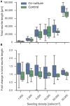
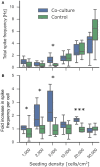
Bottom-up neuroscience aims to engineer well-defined networks of neurons to investigate the functions of the brain. By reducing the complexity of the brain to achievable target questions, such in vitro bioassays better control experimental variables and can serve as a versatile tool for fundamental and pharmacological research. Astrocytes are a cell type critical to neuronal function, and the addition of astrocytes to neuron cultures can improve the quality of in vitro assays. Here, we present cellulose as an astrocyte culture substrate. Astrocytes cultured on the cellulose fiber matrix thrived and formed a dense 3D network. We devised a novel co-culture platform by suspending the easy-to-handle astrocytic paper cultures above neuronal networks of low densities typically needed for bottom-up neuroscience. There was significant improvement in neuronal viability after 5 days in vitro at densities ranging from 50,000 cells/cm2 down to isolated cells at 1,000 cells/cm2. Cultures exhibited spontaneous spiking even at the very low densities, with a significantly greater spike frequency per cell compared to control mono-cultures. Applying the co-culture platform to an engineered network of neurons on a patterned substrate resulted in significantly improved viability and almost doubled the density of live cells. Lastly, the shape of the cellulose substrate can easily be customized to a wide range of culture vessels, making the platform versatile for different applications that will further enable research in bottom-up neuroscience and drug development.
Keywords: astrocyte; cell viability; co-culture; low-density culture; network activity; neurite length; neuron; paper-based.
Figures


Similar articles
-
3-D multi-electrode arrays detect early spontaneous electrophysiological activity in 3-D neuronal-astrocytic co-cultures.Biomed Eng Lett. 2020 Jul 31;10(4):579-591. doi: 10.1007/s13534-020-00166-5. eCollection 2020 Nov. Biomed Eng Lett. 2020. PMID: 33194249 Free PMC article.
-
Calcium imaging, MEA recordings, and immunostaining images dataset of neuron-astrocyte networks in culture under the effect of norepinephrine.Gigascience. 2019 Feb 1;8(2):giy161. doi: 10.1093/gigascience/giy161. Gigascience. 2019. PMID: 30544133 Free PMC article.
-
Palmitoylethanolamide Blunts Amyloid-β42-Induced Astrocyte Activation and Improves Neuronal Survival in Primary Mouse Cortical Astrocyte-Neuron Co-Cultures.J Alzheimers Dis. 2018;61(1):389-399. doi: 10.3233/JAD-170699. J Alzheimers Dis. 2018. PMID: 29154284
-
Cross-talk signals in the CNS: role of neurotrophic and hormonal factors, adhesion molecules and intercellular signaling agents in luteinizing hormone-releasing hormone (LHRH)-astroglial interactive network.Front Biosci. 1997 Mar 1;2:d88-125. doi: 10.2741/a177. Front Biosci. 1997. PMID: 9159216 Review.
-
Local energy on demand: Are 'spontaneous' astrocytic Ca2+-microdomains the regulatory unit for astrocyte-neuron metabolic cooperation?Brain Res Bull. 2018 Jan;136:54-64. doi: 10.1016/j.brainresbull.2017.04.011. Epub 2017 Apr 24. Brain Res Bull. 2018. PMID: 28450076 Review.
Cited by
-
Connectomics of Morphogenetically Engineered Neurons as a Predictor of Functional Integration in the Ischemic Brain.Front Neurol. 2019 Jun 12;10:630. doi: 10.3389/fneur.2019.00630. eCollection 2019. Front Neurol. 2019. PMID: 31249553 Free PMC article. Review.
-
An analytical screening platform to differentiate acute and prolonged exposures of per- and polyfluoroalkyl substances on invasive cellular phenotypes.Toxicol Sci. 2025 Jun 1;205(2):369-379. doi: 10.1093/toxsci/kfaf044. Toxicol Sci. 2025. PMID: 40156146
-
Astrocytes Exhibit a Protective Role in Neuronal Firing Patterns under Chemically Induced Seizures in Neuron-Astrocyte Co-Cultures.Int J Mol Sci. 2021 Nov 25;22(23):12770. doi: 10.3390/ijms222312770. Int J Mol Sci. 2021. PMID: 34884577 Free PMC article.
-
Profiling transcriptomic responses of human stem cell-derived medium spiny neuron-like cells to exogenous phasic and tonic neurotransmitters.Mol Cell Neurosci. 2023 Sep;126:103876. doi: 10.1016/j.mcn.2023.103876. Epub 2023 Jun 28. Mol Cell Neurosci. 2023. PMID: 37385515 Free PMC article.
-
Constructive Neuroengineering of Crossing Multi-Neurite Wiring Using Modifiable Agarose Gel Platforms.Gels. 2025 May 30;11(6):419. doi: 10.3390/gels11060419. Gels. 2025. PMID: 40558718 Free PMC article.
References
-
- Aebersold M. J., Dermutz H., Forró C., Weydert S., Thompson-Steckel G., Vörös J., et al. (2016). Brains on a chip: towards engineered neural networks. TrAC Trends Anal. Chem. 78, 60–69. 10.1016/j.trac.2016.01.025 - DOI
-
- Akram M. S., Daly R., da Cruz Vasconcellos F., Yetisen A. K., Hutchings I., Hall E. A. H. (2015). Applications of paper-based diagnostics, in Lab-on-a-Chip Devices and Micro-Total Analysis Systems (Cham: Springer International Publishing; ), 161–195.
LinkOut - more resources
Full Text Sources
Other Literature Sources

