. 2018 Feb 27:9:134.
doi: 10.3389/fphys.2018.00134. eCollection 2018.

Short-Term Sustained Hypoxia Elevates Basal and Hypoxia-Induced Ventilation but Not the Carotid Body Chemoreceptor Activity in Rats

Affiliations

Short-Term Sustained Hypoxia Elevates Basal and Hypoxia-Induced Ventilation but Not the Carotid Body Chemoreceptor Activity in Rats

Karine C Flor et al. Front Physiol. .

Abstract

Exposure to chronic sustained hypoxia (SH), as experienced in high altitudes, elicits an increase in ventilation, named ventilatory acclimatization to hypoxia (VAH). We previously showed that rats exposed to short-term (24 h) SH exhibit enhanced abdominal expiratory motor activity at rest, accompanied by augmented baseline sympathetic vasoconstrictor activity. In the present study, we investigated whether the respiratory and sympathetic changes elicited by short-term SH are accompanied by carotid body chemoreceptor sensitization. Juvenile male Holtzman rats (60-80 g) were exposed to SH (10% O2 for 24 h) or normoxia (control) to examine basal and hypoxic-induced ventilatory parameters in unanesthetized conditions, as well as the sensory response of carotid body chemoreceptors in artificially perfused in situ preparations. Under resting conditions (normoxia/normocapnia), SH rats (n = 12) exhibited higher baseline respiratory frequency, tidal volume, and minute ventilation compared to controls (n = 11, P < 0.05). SH group also showed greater hypoxia ventilatory response than control group (P < 0.05). The in situ preparations of SH rats (n = 8) exhibited augmented baseline expiratory and sympathetic activities under normocapnia, with additional bursts in abdominal and thoracic sympathetic nerves during late expiratory phase that were not seen in controls (n = 8, P < 0.05). Interestingly, basal and potassium cyanide-induced afferent activity of carotid sinus nerve (CSN) was similar between SH and control rats. Our findings indicate that the maintenance of elevated resting ventilation, baseline sympathetic overactivity, and enhanced ventilatory responses to hypoxia in rats exposed to 24 h of SH are not dependent on increased basal and sensorial activity of carotid body chemoreceptors.

Keywords: active expiration; carotid body; chemoreceptor; hypoxic ventilatory response; sympathetic activity; ventilatory acclimatization.


Figures

Figure 1
Figure 1
Pulmonary ventilation of control and SH rats. Recordings of airflow of control and SH rats, representative from their respective groups, illustrating the ventilatory pattern during baseline and during a new hypoxic episode (7% O2).
Figure 2
Figure 2
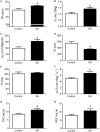
Baseline ventilation in rats after 24 h of sustained hypoxia. Respiratory frequency (Rf, A), tidal volume (VT, B), minute volume (VE, C), expiratory (Te, D), and inspiratory (Ti, E) time, tidal volume to inspiratory time ratio (VT/Ti, F), peak inspiratory flow (PIF, G), and peak expiratory flow (PEF, H) of rats exposed to 24 h of sustained hypoxia (SH, n = 12) or maintained under normoxia (control, n = 11). *Different from control group, P < 0.05.
Figure 3
Figure 3
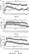
Hypoxic ventilatory response in rats exposed to 24 h of sustained hypoxia. Values of respiratory frequency (Rf, A), tidal volume (VT, B) and minute ventilation (VE, C) of rats exposed to 24 h of sustained hypoxia (SH, n = 12) or maintained under normoxia (control, n = 11) before (time 0), during hypoxia (7% O2 during 20 min, from time 5 to 20) and after the return to normoxia (R). #Different from respective baseline; *Different from control group. P < 0.05.
Figure 4
Figure 4
Baseline and hypoxia-induced changes in body temperature of rats exposed to 24 h of sustained hypoxia. Body temperature at normoxia (time 0), during hypoxia (7% O2, 20 min, gray area) and during the recovery period in rats exposed to 24 h of sustained hypoxia (SH, n = 12) or maintained under normoxia (control, n = 11). #Different from respective baseline (time 0); *Different from control group. P < 0.05.
Figure 5
Figure 5
Baseline respiratory pattern, sympathetic and carotid sinus nerve activity of rats exposed to 24 h of sustained hypoxia. Raw and integrated (∫) recordings of abdominal (AbN), thoracic sympathetic (tSN), phrenic (PN), and carotid sinus (CSN) nerve activities from representative in situ preparations of control (A) and sustained hypoxia-conditioned rats (B). The noise levels of each nerve recording are represented next to the corresponding trace. Dotted lines delineate the inspiratory phase (I), stage 1 of expiration (E1), and stage 2 of expiration (E2).
Figure 6
Figure 6
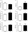
Baseline respiratory and sympathetic parameters of rats exposed to 24 h of sustained hypoxia. Average values of phrenic nerve frequency (PN, A), abdominal mean activity (AbN, B), thoracic sympathetic nerve activity (tSN) during E2 (C), inspiratory (D) and E1 phases (E), and carotid sinus nerve activity (CSN, F) of rats exposed to 24 h of sustained hypoxia (SH, n = 9) or maintained under normoxia (control, n = 8). *Different from control group, P < 0.05.
Figure 7
Figure 7
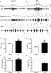
Respiratory and sympathetic responses to stimulation of peripheral chemoreceptors of rats exposed to 24 h of sustained hypoxia. Raw and integrated (∫) recordings of abdominal (AbN), thoracic sympathetic (tSN), phrenic nerve (PN) activities from representative control (A) and sustained hypoxia-conditioned in situ preparations (B), illustrating the responses to peripheral chemoreceptor activation with KCN (0.05%, 50 μL; arrows). (C–F) Average changes in tSN, AbN, PN burst frequency and amplitude, respectively, of rats exposed to 24 h of sustained hypoxia (SH, n = 9) or maintained under normoxia (control, n = 8). *Different from control group, P < 0.05.
Figure 8
Figure 8
Sensory response of carotid body chemoreceptors of rats exposed to 24 h of sustained hypoxia. (A) Raw and integrated (∫) recordings of phrenic (PN) and carotid sinus nerve (CSN) activities from representative preparations of control (A) and sustained hypoxia-conditioned rats (B), illustrating the excitatory response to peripheral chemoreceptor activation with KCN (0.05%, 50 μL; arrow). The noise levels of the carotid sinus nerve recording are represented next to the corresponding trace. (C) Average values of CSN excitatory response to peripheral chemoreceptor activation in rats exposed to 24 h of sustained hypoxia (SH, n = 9) or maintained under normoxia (control, n = 8).

References

    1. Abdala A. P., Rybak I. A., Smith J. C., Paton J. F. (2009). Abdominal expiratory activity in the rat brainstem-spinal cord in situ: patterns, origins and implications for respiratory rhythm generation. J. Physiol. 587, 3539–3559. 10.1113/jphysiol.2008.167502 - DOI - PMC - PubMed
    1. Accorsi-Mendonça D., Almado C. E., Bonagamba L. G., Castania J. A., Moraes D. J., Machado B. H. (2015). Enhanced firing in NTS induced by short-term sustained hypoxia is modulated by glia-neuron interaction. J. Neurosci. 35, 6903–6917. 10.1523/JNEUROSCI.4598-14.2015 - DOI - PMC - PubMed
    1. Angelova P. R., Kasymov V., Christie I., Sheikhbahaei S., Turovsky E., Marina N., et al. . (2015). Functional oxygen sensitivity of astrocytes. J. Neurosci. 35, 10460–10473. 10.1523/JNEUROSCI.0045-15.2015 - DOI - PMC - PubMed
    1. Barnard P., Andronikou S., Pokorski M., Smatresk N., Mokashi A., Lahiri S. (1987). Time-dependent effect of hypoxia on carotid body chemosensory function. J. Appl. Physiol. 63, 685–691. 10.1152/jappl.1987.63.2.685 - DOI - PubMed
    1. Barros R. C., Zimmer M. E., Branco L. G., Milsom W. K. (2001). Hypoxic metabolic response of the golden-mantled ground squirrel. J. Appl. Physiol. 91, 603–612. 10.1152/jappl.2001.91.2.603 - DOI - PubMed

LinkOut - more resources