. 2018 Feb 27:9:327.
doi: 10.3389/fmicb.2018.00327. eCollection 2018.

Lipoteichoic Acid Inhibits Staphylococcus aureus Biofilm Formation

Affiliations

Lipoteichoic Acid Inhibits Staphylococcus aureus Biofilm Formation

Ki Bum Ahn et al. Front Microbiol. .

Abstract

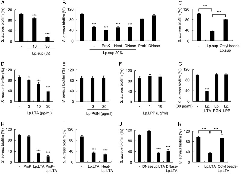
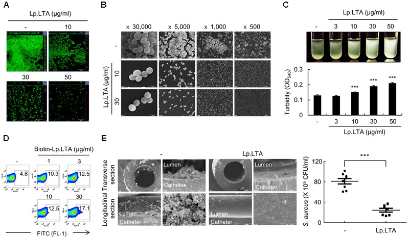
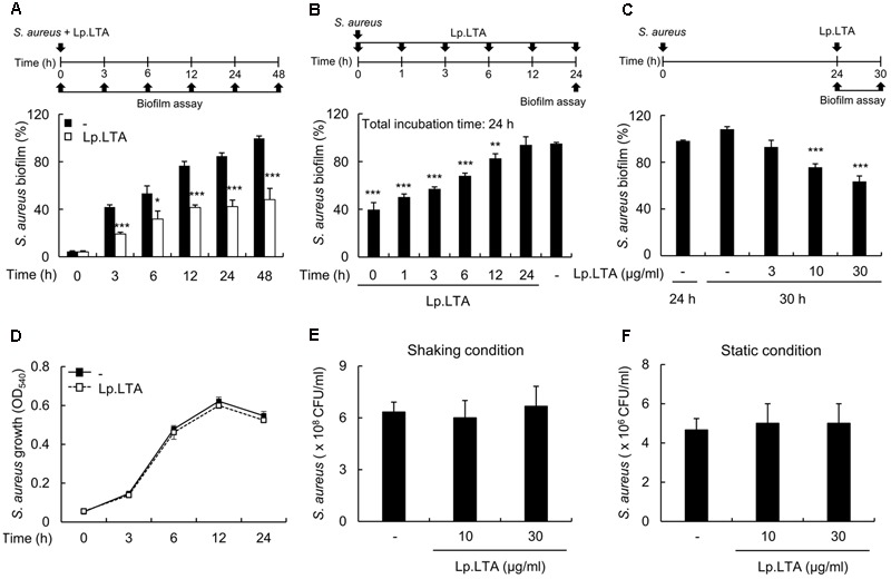
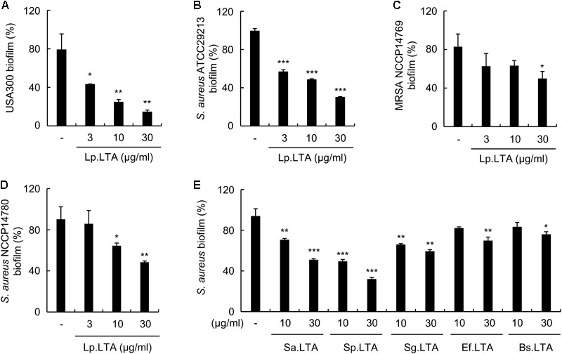
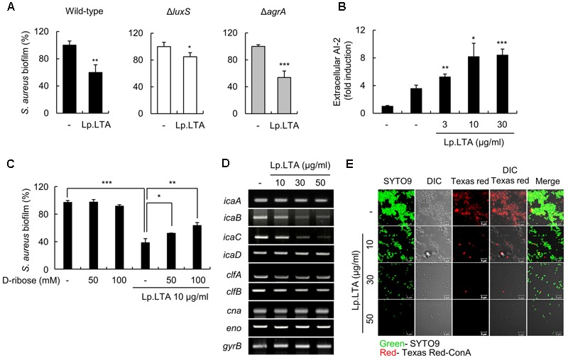
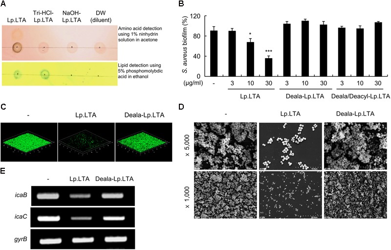
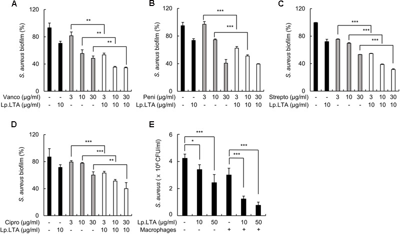
A biofilm is an aggregate of microorganisms in which cells adhere to biological or non-biological surfaces and is responsible for various infectious diseases. Infections caused by Staphylococcus aureus, including pneumonia, endocarditis, and osteomyelitis, are often associated with colonization and biofilm formation. Although lipoteichoic acid (LTA) is involved in biofilm formation, the specific role of LTA is not clearly understood. In this study, we demonstrated that LTA released from Lactobacillus plantarum could inhibit S. aureus biofilm formation and aggregation without affecting the growth of S. aureus in various in vitro and in vivo models. L. plantarum LTA (Lp.LTA) also inhibited biofilm formation of S. aureus clinical isolates, including a methicillin-resistant strain. Remarkably, Lp.LTA not only interfered with S. aureus biofilm formation, but it also disrupted a pre-formed biofilm. Mechanism studies demonstrated that Lp.LTA inhibited expression of the ica-operon, which is responsible for the production of poly-N-acetylglucosamine, a key molecule required for S. aureus biofilm development. Lp.LTA increased the release of autoinducer-2 from S. aureus, which contributed to the inhibition of S. aureus biofilm formation. Moreover, Lp.LTA treatment enhanced susceptibility of the biofilm to various antibiotics and to macrophages. Interestingly, Lp.LTA without D-alanine moieties was not able to inhibit biofilm formation by S. aureus. In conclusion, the present study suggests that LTA can inhibit S. aureus biofilm formation, and therefore could be applied for preventing and/or treating infectious diseases caused by S. aureus biofilms.

Keywords: Lactobacillus plantarum; Staphylococcus aureus; biofilm formation; infectious diseases; lipoteichoic acid.


Figures

FIGURE 1
FIGURE 1
L. plantarum LTA has inhibitory effects on S. aureus biofilm formation. S. aureus (5 × 10⁷ CFU/ml) was grown on polystyrene plates at 37°C for 24 h in the presence of (A) L. plantarum culture supernatants (Lp.sup) (10 or 30%), (B) 50 μg/ml of proteinase K (ProK)-treated Lp.sup (20%), heat-treated Lp.sup (20%) for 10 min at 100°C, or 50 μg/ml of DNase I (DNase)-treated Lp.sup (20%), (C) octyl-sepharose bead-treated Lp.sup (20%), (D) L. plantarum LTA (Lp.LTA) (3, 10, or 30 μg/ml), (E) L. plantarum peptidoglycan (Lp.PGN) (3 or 30 μg/ml), (F) L. plantarum lipoprotein (Lp.LPP) (1 or 10 μg/ml), (G) S. aureus (5 × 10⁷ CFU/ml) was grown on sterile glass coverslips at 37°C for 24 h in the presence of Lp.LTA (30 μg/ml), Lp.PGN (30 μg/ml), or Lp.LPP (30 μg/ml). S. aureus (5 × 10⁷ CFU/ml) was grown on polystyrene plates at 37°C for 24 h in the presence of 30 μg/ml of Lp.LTA or (H) 50 μg/ml of proteinase K-treated Lp.LTA (30 μg/ml), (I) heat-treated Lp.LTA (30 μg/ml), (J) 50 μg/ml of DNase I-treated Lp.LTA (30 μg/ml), or (K) octyl-sepharose bead-treated Lp.LTA (30 μg/ml). The extent of biofilm formation was determined via crystal violet assay. Data are the mean values ± SD of triplicate samples. Asterisks indicate significant induction compared with the non-treatment group (P < 0.05 and ∗∗∗P < 0.001).
FIGURE 2
FIGURE 2
L. plantarum LTA inhibits biofilm formation and aggregation of S. aureus in vitro and in vivo. (A,B) S. aureus (5 × 10⁷ CFU/ml) was grown on polystyrene plates at 37°C for 24 h in the absence or presence of Lp.LTA (10, 30, or 50 μg/ml). Lp.LTA-treated S. aureus biofilms were visualized (A) by confocal laser scanning microscopy (green from SYTO9 and red from propidium iodide) or (B) by scanning electron microscopy (Magnification: ×500, ×1,000, ×5,000, or ×30,000). (C) S. aureus (2 × 10⁸ CFU/ml) was grown for 24 h in the absence or presence of 3, 10, 30, or 50 μg/ml of Lp.LTA. Aggregation of S. aureus was determined by optical density (O.D.) at 540 nm. Data are the mean values ± SD of triplicate samples. (D) S. aureus (5 × 10⁷ CFU/ml) was treated with 1, 3, 10, or 30 μg/ml of biotinylated Lp.LTA (biotin-Lp.LTA) for 1 h. Binding of biotin-Lp.LTA with S. aureus was detected by streptavidin-FITC and analyzed via flow cytometry. The percentage of biotin-Lp.LTA-positive S. aureus is shown in each histogram. One of three similar results is shown. (E) C57BL/6 mice were implanted with S. aureus-loaded catheter in the presence or absence of Lp.LTA. After 3 days, the mice were sacrificed to harvest the catheters. The catheters were sectioned to obtain transverse rings and longitudinal strips, which were subject to SEM analysis (Magnification: ×100, ×2,000, ×5,000, and ×100, clockwise in each treatment group), and the remaining biofilms on the catheters were measured by counting colony forming units. Eight and seven mice were used in the non-treatment and LpLTA-treatment groups, respectively. Asterisks indicate significant induction compared with the non-treatment group (∗∗∗P < 0.001).
FIGURE 3
FIGURE 3
L. plantarum LTA inhibits biofilm development at early and late stages, and even disrupts the pre-formed biofilm. (A) S. aureus (1 × 10⁷ CFU/ml) was grown on polystyrene plates at 37°C for 3, 6, 12, 24, or 48 h in the presence or absence of Lp.LTA (30 μg/ml). (B) S. aureus (5 × 10⁷ CFU/ml) was grown on polystyrene plates at 37°C for 1, 3, 6, or 12 h, followed by treatment with Lp.LTA (30 μg/ml) at 37°C for up to 24 h. (C) S. aureus (5 × 10⁷ CFU/ml) was grown on polystyrene plates at 37°C for 24 h, and then supernatant containing planktonic bacteria was removed. Pre-formed biofilm was treated with Lp.LTA (3, 10, or 30 μg/ml) and further incubated at 37°C for 6 h. Biofilm formation was determined by a crystal violet assay. (D) S. aureus (1 × 10⁷ CFU/ml) was grown under shaking conditions for 3, 6, 12, or 24 h in the presence or absence of Lp.LTA (30 μg/ml). The growth of S. aureus was determined by O.D. at 540 nm. (E,F) S. aureus (5 × 10⁷ CFU/ml) was grown in the presence or absence of Lp.LTA (10 or 30 μg/ml) for 24 h under (E) shaking or (F) static culture condition. Bacterial viability was measured by counting colony forming units. Data are the mean values ± SD of triplicate samples. Asterisks indicate significant differences compared with the non-treatment group (P < 0.05, ∗∗P < 0.01, and ∗∗∗P < 0.001).
FIGURE 4
FIGURE 4
L. plantarum LTA inhibits biofilm formation in various strains of S. aureus, and purified LTAs from various Gram-positive bacteria also have an inhibitory effect. (A) S. aureus USA300, (B) ATCC 29213, (C) NCCP14769, or (D) NCCP14780 were treated with 3, 10, or 30 μg/ml of Lp.LTA for 24 h. (E) S. aureus (5 × 10⁷ CFU/ml) was grown on polystyrene plates at 37°C for 24 h in the presence of purified LTA from S. aureus, S. pneumoniae, S. gordonii, E. faecalis, or B. subtilis. Biofilm formation was determined via crystal violet assay. Data are the mean values ± SD of triplicate samples. Asterisks indicate significant inhibition compared with the non-treatment group (P < 0.05, ∗∗P < 0.01, and ∗∗∗P < 0.001).
FIGURE 5
FIGURE 5
L. plantarum LTA induces AI-2 release and down-regulates ica gene expression and exopolysaccharide production in S. aureus. (A) S. aureus wild-type (1 × 10⁷ CFU/ml), an AI-2-deficient strain (ΔluxS) (1 × 10⁷ CFU/ml), and an AIP-deficient strain (ΔagrA) (1 × 10⁷ CFU/ml) were grown in the absence or presence of Lp.LTA (10 μg/ml) at 37°C for 24 h. (B) S. aureus (1 × 10⁷ CFU/ml) was grown in the absence or presence of Lp.LTA (3, 10, or 30 μg/ml) on polystyrene plates at 37°C for 24 h. The culture supernatants were collected to determine AI-2 release using a bioluminescent bacterial reporter strain, Vibrio harveyi BB170. (C) S. aureus (5 × 10⁷ CFU/ml) was grown on polystyrene plates at 37°C for 24 h in the absence or presence of Lp.LTA (10 μg/ml) with 50 or 100 mM of D-ribose. Biofilm formation was determined via crystal violet assay. Data are the mean values ± SD of triplicate samples. (D) S. aureus (1 × 10⁸ CFU/ml) was treated with 0, 10, 30, or 50 μg/ml of Lp.LTA for 3 h. Total RNA was isolated, and the mRNA expression of icaA, icaB, icaC, icaD, clfA, clfB, cna, eno, and gyrB was examined by RT-PCR. (E) S. aureus (5 × 10⁷ CFU/ml) was grown at 37°C for 24 h in the presence of Lp.LTA (0, 10, 30, or 50 μg/ml). Then, S. aureus biofilm formation and exopolysaccharide production were examined with SYTO9 and Texas Red-ConA staining, respectively, followed by analysis using confocal microscopy (green from SYTO9 and red from Texas Red-ConA). Asterisks indicate significant induction compared with the non-treatment group (P < 0.05, ∗∗P < 0.01, and ∗∗∗P < 0.001).
FIGURE 6
FIGURE 6
D-Alanine moieties of Lp.LTA are critical for the inhibitory effect on S. aureus biofilm formation. (A) Dealanylated (Deala)-Lp.LTA and dealanylated/deacylated (Deala/Deacyl)-Lp.LTA were prepared by treatment of Lp.LTA with Tris-HCl (0.1 M, pH 8.5) or NaOH (0.5 N, pH 12), respectively. Then, D-alanine and acyl chain moieties of Lp.LTA were detected by 1% ninhydrin solution and 5% phosphomolybdic acid, respectively. (B) S. aureus (5 × 10⁷ CFU/ml) was grown on polystyrene plates at 37°C for 24 h in the presence of 0, 3, 10, or 30 μg/ml of Lp.LTA, Deala-Lp.LTA, or Deala/Deacyl-Lp.LTA. Biofilm formation was determined by a crystal violet assay. Data are the mean values ± SD of triplicate samples. (C) S. aureus (5 × 10⁷ CFU/ml) was grown on glass-bottom dishes at 37°C for 24 h in the absence or presence of 30 μg/ml of Lp.LTA or Deala-Lp.LTA. S. aureus biofilms were visualized by confocal laser scanning microscopy (green from SYTO9 and red from propidium iodide). (D) S. aureus (5 × 10⁷ CFU/ml) was grown at 37°C for 24 h in the absence or presence of 30 μg/ml of Lp.LTA or Deala-Lp.LTA. S. aureus biofilms were analyzed by scanning electron microscopy (magnification: ×1,000 or ×5,000). (E) S. aureus (1 × 10⁸ CFU/ml) was treated with 30 μg/ml of Lp.LTA or Deala-Lp.LTA for 3 h. Total RNA was isolated, and the expression of icaB, icaC, and gyrB mRNA was examined by RT-PCR. Asterisks indicate significant induction compared with the non-treatment group (P < 0.05 and ∗∗∗P < 0.001).
FIGURE 7
FIGURE 7
L. plantarum LTA increases the susceptibility of S. aureus biofilms to antibiotics and macrophages. S. aureus (5 × 10⁷ CFU/ml) was grown on polystyrene plates with 10 μg/ml of Lp.LTA at 37°C for 24 h, and further incubated for 6 h in the absence or presence of 3, 10, or 30 μg/ml of antibiotics that target (A,B) cell wall synthesis (vancomycin and penicillin), (C) protein synthesis (streptomycin), or (D) nucleic acid synthesis (ciprofloxacin). The extent of biofilm formation was determined by a crystal violet assay. Data are the mean values ± SD of triplicate samples. (E) S. aureus (5 × 10⁷ CFU/ml) was grown on sterile glass coverslips at 37°C for 24 h in the presence or absence of Lp.LTA (10 or 50 μg/ml) and treated with RAW 264.7 cells (1 × 10⁶ cells/ml) at 37°C for 2 h. Remaining biofilms were measured by counting colony forming units. Data are the mean values ± SD of triplicate samples. Asterisks indicate significant induction compared with the non-treatment group or the macrophages alone-treatment group (P < 0.05, ∗∗P < 0.01, and ∗∗∗P < 0.001).

References

    1. Atshan S. S., Nor Shamsudin M., Sekawi Z., Lung L. T., Hamat R. A., Karunanidhi A., et al. (2012). Prevalence of adhesion and regulation of biofilm-related genes in different clones of Staphylococcus aureus. J. Biomed. Biotechnol. 2012:976972. 10.1155/2012/976972 - DOI - PMC - PubMed
    1. Baik J. E., Jang K. S., Kang S. S., Yun C. H., Lee K., Kim B. G., et al. (2011). Calcium hydroxide inactivates lipoteichoic acid from Enterococcus faecalis through deacylation of the lipid moiety. J. Endod. 37 191–196. 10.1016/j.joen.2010.11.007 - DOI - PubMed
    1. Baik J. E., Ryu Y. H., Han J. Y., Im J., Kum K. Y., Yun C. H., et al. (2008). Lipoteichoic acid partially contributes to the inflammatory responses to Enterococcus faecalis. J. Endod. 34 975–982. 10.1016/j.joen.2008.05.005 - DOI - PubMed
    1. Bassler B. L., Wright M., Showalter R. E., Silverman M. R. (1993). Intercellular signalling in Vibrio harveyi: sequence and function of genes regulating expression of luminescence. Mol. Microbiol. 9 773–786. 10.1111/j.1365-2958.1993.tb01737.x - DOI - PubMed
    1. Biswas R., Voggu L., Simon U. K., Hentschel P., Thumm G., Gotz F. (2006). Activity of the major staphylococcal autolysin Atl. FEMS Microbiol. Lett. 259 260–268. 10.1111/j.1574-6968.2006.00281.x - DOI - PubMed

LinkOut - more resources