

Selenium-binding protein 1 is down-regulated in malignant melanoma
- PMID: 29535818
- PMCID: PMC5828193
- DOI: 10.18632/oncotarget.23853
Selenium-binding protein 1 is down-regulated in malignant melanoma
Abstract
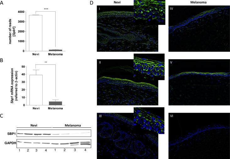
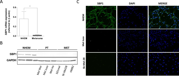
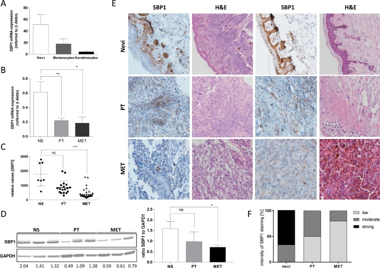
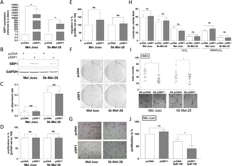
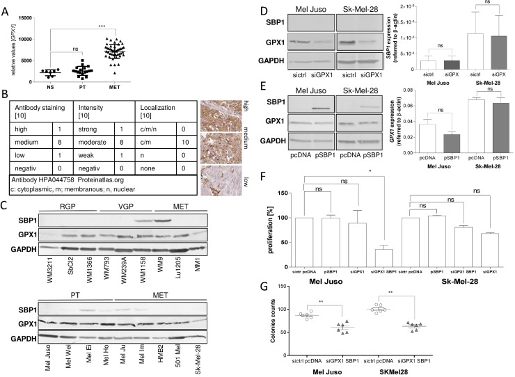
Selenium-binding protein 1 (SELENBP1) expression is reduced in various epithelial cancer entities compared to corresponding normal tissue and has already been described as a tumor suppressor involved in the regulation of cell proliferation, senescence, migration and apoptosis. We identified SELENBP1 to be down-regulated in cutaneous melanoma, a malignant cancer of pigment-producing melanocytes in the skin, which leads to the assumption that SELENBP1 also functions as tumor suppressor in the skin, as shown by others e.g. for prostate or lung carcinoma. However, in vitro analyses indicate that SELENBP1 re-expression in human melanoma cell lines has no impact on cell proliferation, migration or tube formation of the tumor cells themselves when compared to control-transfected cells. Interestingly, supernatant taken from melanoma cell lines transfected with a SELENBP1 re-expression plasmid led to suppression of vessel formation of HMEC cells. Furthermore, SELENBP1 re-expression alters the sensitivity of melanoma cells for Vemurafenib treatment. The data also hint to a functional interaction of SELENBP1 with GPX1 (Glutathione peroxidase 1). Low SELENBP1 mRNA levels correlate inversely with GPX1 expression in melanoma. The re-expression of SELENBP1 combined with down-regulation of GPX1 expression led to reduction of the proliferation of melanoma cells. In summary, SELENBP1 influences the tumor microenvironment and SELENBP1 action is functionally influenced by GPX1.
Keywords: Grm1 mouse model; glutathione peroxidase 1 (GPX1); malignant melanoma; selenium-binding protein 1 (SELENBP1).
Conflict of interest statement
CONFLICTS OF INTEREST The authors declare no conflicts of interest.
Figures
Similar articles
-
Selenium-binding protein 1 inhibits malignant progression and induces apoptosis via distinct mechanisms in non-small cell lung cancer.Cancer Med. 2023 Aug;12(16):17149-17170. doi: 10.1002/cam4.6309. Epub 2023 Aug 22. Cancer Med. 2023. PMID: 37606338 Free PMC article.
-
Reduced selenium-binding protein 1 in breast cancer correlates with poor survival and resistance to the anti-proliferative effects of selenium.PLoS One. 2013 May 21;8(5):e63702. doi: 10.1371/journal.pone.0063702. Print 2013. PLoS One. 2013. PMID: 23704933 Free PMC article.
-
Selenium binding protein 1 in ovarian cancer.Int J Cancer. 2006 May 15;118(10):2433-40. doi: 10.1002/ijc.21671. Int J Cancer. 2006. PMID: 16380993
-
The role of SELENBP1 and its epigenetic regulation in carcinogenic progression.Front Genet. 2022 Nov 1;13:1027726. doi: 10.3389/fgene.2022.1027726. eCollection 2022. Front Genet. 2022. PMID: 36386843 Free PMC article. Review.
-
Apoptosis and pathogenesis of melanoma and nonmelanoma skin cancer.Adv Exp Med Biol. 2008;624:283-95. doi: 10.1007/978-0-387-77574-6_22. Adv Exp Med Biol. 2008. PMID: 18348464 Review.
Cited by
-
Loss of CYLD accelerates melanoma development and progression in the Tg(Grm1) melanoma mouse model.Oncogenesis. 2019 Oct 7;8(10):56. doi: 10.1038/s41389-019-0169-4. Oncogenesis. 2019. PMID: 31591386 Free PMC article.
-
Serum Selenium Level and 10-Year Survival after Melanoma.Biomedicines. 2021 Aug 11;9(8):991. doi: 10.3390/biomedicines9080991. Biomedicines. 2021. PMID: 34440195 Free PMC article.
-
Selenium-Binding Protein 1 (SELENBP1) as Biomarker for Adverse Clinical Outcome After Traumatic Spinal Cord Injury.Front Neurosci. 2021 May 28;15:680240. doi: 10.3389/fnins.2021.680240. eCollection 2021. Front Neurosci. 2021. PMID: 34140879 Free PMC article.
-
Construction and Validation of a 9-Gene Signature for Predicting Prognosis in Stage III Clear Cell Renal Cell Carcinoma.Front Oncol. 2019 Mar 19;9:152. doi: 10.3389/fonc.2019.00152. eCollection 2019. Front Oncol. 2019. PMID: 30941304 Free PMC article.
-
A component overlapping attribute clustering (COAC) algorithm for single-cell RNA sequencing data analysis and potential pathobiological implications.PLoS Comput Biol. 2019 Feb 19;15(2):e1006772. doi: 10.1371/journal.pcbi.1006772. eCollection 2019 Feb. PLoS Comput Biol. 2019. PMID: 30779739 Free PMC article.
References
-
- Martín García E, Arias-Santiago S, Serrano-Ortega S, Buendía-Eisman A. [Changes in the Incidence of Skin and Lip Cancer Between 1978 and 2007.] [Article in Spanish] Actas Dermosifiliogr. 2017;108:335–45. - PubMed
-
- Pollock PM, Cohen-Solal K, Sood R, Namkoong J, Martino JJ, Koganti A, Zhu H, Robbins C, Makalowska I, Shin SS, Marin Y, Roberts KG, Yudt LM, et al. Melanoma mouse model implicates metabotropic glutamate signaling in melanocytic neoplasia. Nat Genet. 2003;34:108–112. - PubMed
-
- Schiffner S, Braunger BM, de Jel MM, Coupland SE, Tamm ER, Bosserhoff AK. Tg(Grm1) transgenic mice: a murine model that mimics spontaneous uveal melanoma in humans? Exp Eye Res. 2014;127:59–68. - PubMed
-
- Namkoong J, Shin SS, Lee HJ, Marin YE, Wall BA, Goydos JS, Chen S. Metabotropic glutamate receptor 1 and glutamate signaling in human melanoma. Cancer Res. 2007;67:2298–2305. - PubMed
LinkOut - more resources
Full Text Sources
Other Literature Sources
Research Materials
Miscellaneous
