. 2018 Jan 31;9(12):10665-10680.
doi: 10.18632/oncotarget.24366. eCollection 2018 Feb 13.

Innovative methodology for the identification of soluble biomarkers in fresh tissues

Affiliations

Innovative methodology for the identification of soluble biomarkers in fresh tissues

Brunella Costanza et al. Oncotarget. .

Abstract

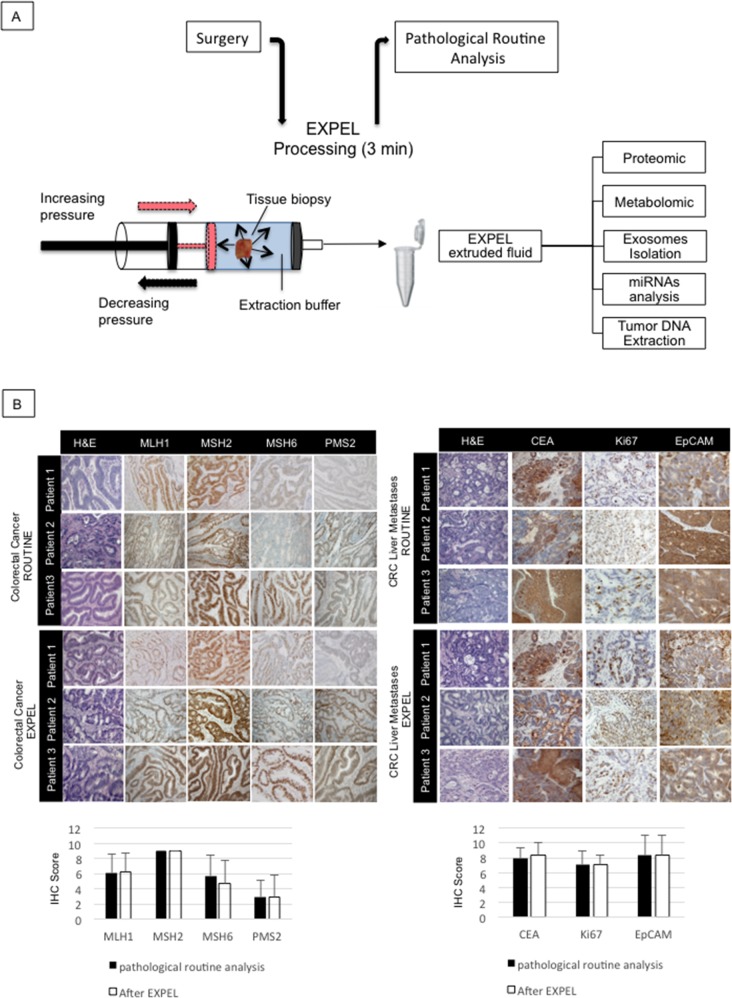
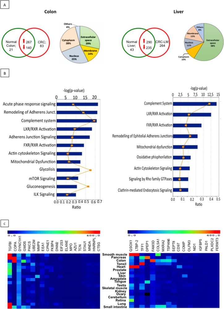
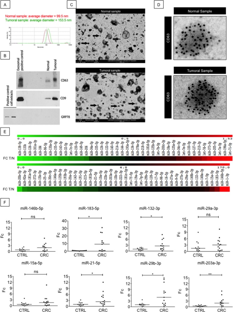
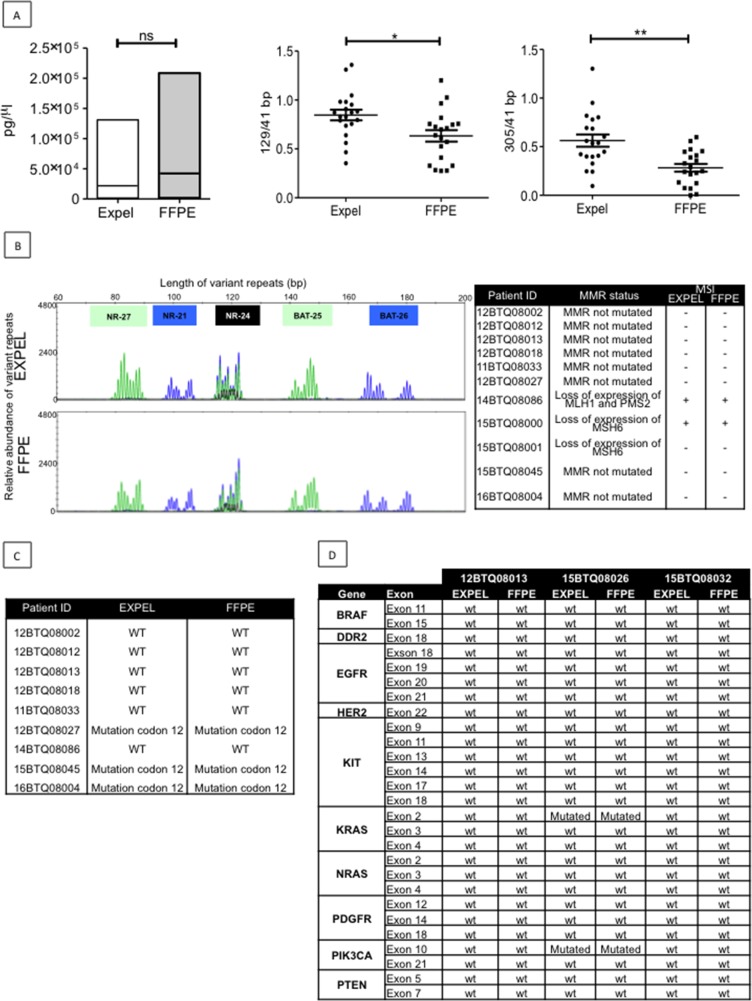
The identification of diagnostic and prognostic biomarkers from early lesions, measurable in liquid biopsies remains a major challenge, particularly in oncology. Fresh human material of high quality is required for biomarker discovery but is often not available when it is totally required for clinical pathology investigation. Hence, all OMICs studies are done on residual and less clinically relevant biological samples. Here after, we present an innovative, simple, and non-destructive, procedure named EXPEL that uses rapid, pressure-assisted, interstitial fluid extrusion, preserving the specimen for full routine clinical pathology investigation. In the meantime, the technique allows a comprehensive OMICs analysis (proteins, metabolites, miRNAs and DNA). As proof of concept, we have applied EXPEL on freshly collected human colorectal cancer and liver metastases tissues. We demonstrate that the procedure efficiently allows the extraction, within a few minutes, of a wide variety of biomolecules holding diagnostic and prognostic potential while keeping both tissue morphology and antigenicity unaltered. Our method enables, for the first time, both clinicians and scientists to explore identical clinical material regardless of its origin and size, which has a major positive impact on translation to the clinic.

Keywords: biomarkers; metabolomic; miRNAs; proteomic; tDNA.


Conflict of interest statement

CONFLICTS OF INTEREST The authors declare no conflicts of interest.

Figures

Figure 1
Figure 1. EXPEL method does not alter tissue morphology and antigenicity
(A) Schematic overview of EXPEL workflow. Diagram of standard tissue processing from the surgery room to pathologists. EXPEL extruded fluid obtained within 3 minutes from a tissue sample, is prepared for the indicated applications or stored at −20°C. (B) Colorectal primary tumors (n=10, left panel) and liver metastases (n=10, right panel) were subjected to clinical routine analysis and EXPEL method followed by hematoxylin/eosin (H&E) staining and immunolabelling of the indicated markers (representative pictures are shown for 3 patients). The quantitative evaluation for each marker was assessed as outlined in the Material and Methods section. The error bars indicate standard deviation of means. Images of representative fields were taken at 100× magnification.
Figure 2
Figure 2. Proteomic analysis of EXPEL extruded fluid identifies potential cancer biomarkers
(A) Absolute numbers of proteins, identified after proteomic analysis of EXPEL extruded fluids, in at least 4 out of 7 for CRC (upper, left panel) and 3 out of 6 for CRC-LM (upper right, panel) replicates. Pie charts indicating the predicted sub-cellular localization of identified proteins for CRC (on left side) and CRC-LM (on right side) are shown. (B) Significantly altered canonical pathways of cancer up-regulated and uniquely expressed proteins analyzed by the IPA software using IPA Core Analysis for colon (on the left) and liver (on the right) EXPEL extruded fluids. The canonical pathways are shown along the y-axis of the bar chart. The x-axis indicates the statistical significance (on the upper part, calculated using the right-tailed Fisher exact test. The P value indicates which biologic annotations are significantly associated with the input molecules relative to all functionally characterized mammalian molecules. “Ratio” (differential yellow line and markers) refers to the number of molecules from the dataset that map to the pathway listed divided by the total number of molecules that map to the canonical pathway from within the IPA knowledgebase. (C) Normal tissue gene expression of potential biomarkers candidates discovered from colorectal cancer (left panel) and liver metastases (right panel). mRNA expressions were assessed using BioGPS public database.
Figure 3
Figure 3. EXPEL extruded fluid contains exosomes and miRNAs readily detectable in patients sera
(A) Dynamic light scattering of isolated exosomes from normal and tumoral samples revealed that the average diameter of the vesicles is in the expected size range, a representative image of three independent experiment is shown. (B) Western blot validation of exosomal preparations. CD9 and CD63 are used as positive control for exosomes isolation whereas GRP78 is employed as control to exclude the presence of contaminating organelles. (C) Electron microscopy revealed the presence of exosomes (black arrows) in EXPEL extruded fluid. A representative image of two independent experiments is shown. (D) A representative picture of anti-CD63 immunogold labeling evidencing specific staining for exosomal vesicles in normal and tumoral samples. (E) miRNome analysis of 84 different miRNAs in EXPEL extruded fluid. The amplified miRNAs are shown in heat maps indicating fold-change differences in tumoral samples versus their normal counterpart (FC T/N). Samples analyzed correspond to a pool of 3 EXPEL extruded fluids from CRC and 3 of their matched normal counterparts. (F) Validation of the indicated miRNAs in serum samples of healthy donors (CTRL, n=11) and CRC patients (n=13). Error bars indicate standard deviation of means. Statistical analysis was performed using Whitney U test (* p<0.05, ** p<0.01 and *** p<0.001).
Figure 4
Figure 4. EXPEL extruded fluid contains high quality tumor DNA (tDNA) that is exploitable for genetic profiling
(A) Left panel, equivalent yield of tDNA was obtained from FFPE sections and matched EXPEL fluids of CRC primary lesions (n=10) and CRC-liver metastatic lesions (n=10). The quality of the DNA was assessed on the same extracts. The ratios of long 129bp (middle panel) and 305bp (right panel) to short amplified fragments (41bp) indicated that EXPEL tDNA presented with significantly higher amounts of long fragments in comparison with the FFPE tDNA. Dot plots show the mean ± SEM. Statistical significance was calculated using Wilcoxon paired test (* p<0.05, ** p<0.01 and *** p<0.001). (B) PCR microsatellite instability (MSI) analysis at 5 loci (NR-27, NR-21, NR- 24, BAT-25 and BAT-26) of a representative CRC primary tumor shows similar electropherograms for both FFPE and EXPEL tDNA extracts (left panel). Right panel summarizes MSI analysis results obtained for a subset of 11 tumor samples. The corresponding MMR status, based on routine IHC detection of MMR genes (MLH1, MSH2, MSH6, PMS2), is given. (C) Concordant KRAS mutational status (codon 12) detected using pyrosequencing on FFPE and EXPEL tDNAs isolated from CRC primary lesions and CRC-liver metastatic lesions. (D) Next-generation sequencing (NGS) technique used for the detection of 10 cancer related genetic alterations showed identical results on FFPE and EXPEL tDNAs isolated from CRC and CRC-LM lesions.
Figure 5
Figure 5. Metabolomic profile of EXPEL extruded fluids from CRC and CRC-LM
(A) Pearson correlation clustering of general metabolites quantified in the EXPEL extruded fluids from CRC (on the left, n=13) and CRC-LM (on the right, n=9). The individual values are relative quantification ratios of CRC and CRC-LM versus their normal colon and liver adjacent tissues. (B) Top 10 up- and down- modulated metabolites are shown for CRC (on the left) and CRC-LM (on the right) EXPEL extruded fluids. Error bars indicate standard error of means.

References

    1. Kulasingam V, Diamandis EP. Strategies for discovering novel cancer biomarkers through utilization of emerging technologies. Nat Clin Pract Oncol. 2008;5:588–99. https://doi.org/10.1038/ncponc1187 - DOI - PubMed
    1. McDermott JE, Wang J, Mitchell H, Webb-Robertson BJ, Hafen R, Ramey J, Rodland KD. Challenges in biomarker discovery: combining expert insights with statistical analysis of complex omics data. Expert Opin Med Diagn. 2013;7:37–51. https://doi.org/10.1517/17530059.2012.718329 - DOI - PMC - PubMed
    1. Celis JE, Gromov P, Cabezon T, Moreira JM, Ambartsumian N, Sandelin K, Rank F, Gromova I. Proteomic characterization of the interstitial fluid perfusing the breast tumor microenvironment: a novel resource for biomarker and therapeutic target discovery. Mol Cell Proteomics. 2004;3:327–44. https://doi.org/10.1074/mcp.M400009-MCP200 - DOI - PubMed
    1. Brown KJ, Formolo CA, Seol H, Marathi RL, Duguez S, An E, Pillai D, Nazarian J, Rood BR, Hathout Y. Advances in the proteomic investigation of the cell secretome. Expert Rev Proteomics. 2012;9:337–45. https://doi.org/10.1586/epr.12.21 - DOI - PMC - PubMed
    1. Gronborg M, Kristiansen TZ, Iwahori A, Chang R, Reddy R, Sato N, Molina H, Jensen ON, Hruban RH, Goggins MG, Maitra A, Pandey A. Biomarker discovery from pancreatic cancer secretome using a differential proteomic approach. Mol Cell Proteomics. 2006;5:157–71. https://doi.org/10.1074/mcp.M500178-MCP200 - DOI - PubMed