The Combined Use of Intraluminal and Intrasaccular Flow Diversion for the Treatment of Intracranial Aneurysms: Report of 25 Cases
- PMID: 29535895
- PMCID: PMC5847887
- DOI: 10.5469/neuroint.2018.13.1.20
The Combined Use of Intraluminal and Intrasaccular Flow Diversion for the Treatment of Intracranial Aneurysms: Report of 25 Cases
Abstract
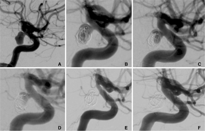
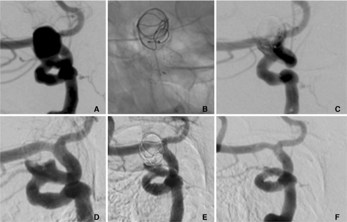
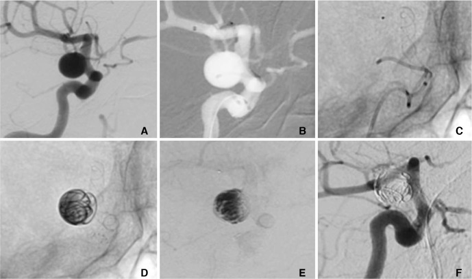
Purpose: The Medina Embolic Device (MED) is a new intrasaccular device with promising early results. Previously we documented our initial experience of this device both alone and in combination with other devices including flow diverter stents (FDS). We sought to determine the effect of the MED + FDS strategy for the treatment of selected aneurysms.
Materials and methods: We performed a retrospective analysis of prospectively collected data to identify all patients with aneurysms treated using both the MED and intraluminal FDS. We present our technical success rate, early and mid-term angiographic follow-up, and clinical outcome data.
Results: We identified 25 non-consecutive patients. The treatment was staged in 9 patients and in a single session 16 patients. The average age was 61±12.8 years (range 40-82). The average fundus height was 11±3.6 mm and average fundus width was 10.1±3.4 mm. In the staged cohort (n=9) at delayed angiography (mean 10 mths) 8 aneurysms (89%) showed complete exclusion (mRRC 1) and in one patient there was a parent vessel occlusion. In the simultaneous cohort delayed angiography (n=10, mean 8.1 months) demonstrated complete occlusion (mRRC 1) in 6 aneurysms (60%), 3 neck remnants (mRRC 2) (30%) and 1 patient (10%) showed persistent aneurysmal filling (mRRC 3a). There were 5 complications with permanent morbidity (mRS >2) in two patients. There were no mortalities.
Conclusion: The MED can be successfully used in combination with intraluminal FDS and in selected aneurysms this may represent an alternative to FDS and adjunctive coiling.
Keywords: Intracranial aneurysm; Medina embolization device; flow diversion.
Conflict of interest statement
P. Bhogal and M. AlMatter serve as proctors and consultants for phenox. H. Henkes is a co-founder and share-holder of phenox. The other authors report no conflict of interest.
Figures
Similar articles
-
The Medina Embolic Device: Karolinska experience.Interv Neuroradiol. 2018 Feb;24(1):4-13. doi: 10.1177/1591019917733125. Epub 2017 Sep 28. Interv Neuroradiol. 2018. PMID: 28956513 Free PMC article.
-
The Medina Embolic Device: early clinical experience from a single center.J Neurointerv Surg. 2017 Jan;9(1):77-87. doi: 10.1136/neurintsurg-2016-012539. Epub 2016 Aug 2. J Neurointerv Surg. 2017. PMID: 27484746 Free PMC article.
-
Medina embolization device for the treatment of intracranial aneurysms: 18 months' angiographic results.J Neurointerv Surg. 2019 May;11(5):516-522. doi: 10.1136/neurintsurg-2018-014110. Epub 2018 Nov 24. J Neurointerv Surg. 2019. PMID: 30472677
-
Treatment of posterior circulation non-saccular aneurysms with flow diverters: a single-center experience and review of 56 patients.J Neurointerv Surg. 2017 May;9(5):471-481. doi: 10.1136/neurintsurg-2016-012781. Epub 2016 Nov 11. J Neurointerv Surg. 2017. PMID: 27836994 Free PMC article. Review.
-
Acutely Ruptured Intracranial Aneurysms Treated with Flow-Diverter Stents: A Systematic Review and Meta-Analysis.AJNR Am J Neuroradiol. 2018 Sep;39(9):1669-1675. doi: 10.3174/ajnr.A5730. Epub 2018 Jul 26. AJNR Am J Neuroradiol. 2018. PMID: 30049721 Free PMC article.
Cited by
-
Placement of a Stent within a Flow Diverter Improves Aneurysm Occlusion Rates.AJNR Am J Neuroradiol. 2019 Nov;40(11):1932-1938. doi: 10.3174/ajnr.A6237. Epub 2019 Oct 3. AJNR Am J Neuroradiol. 2019. PMID: 31582390 Free PMC article.
-
Early Complete Obliteration of Recurrent Large Basilar Aneurysm by Combined Use of Additional Woven EndoBridge Device and Flow Diverter: A Case Report.Neurointervention. 2025 Jul;20(2):94-98. doi: 10.5469/neuroint.2025.00318. Epub 2025 Jun 2. Neurointervention. 2025. PMID: 40451612 Free PMC article.
-
Comaneci plus Balloon-assisted Embolization of Ruptured Wide-necked Cerebral Aneurysms.Clin Neuroradiol. 2022 Sep;32(3):773-782. doi: 10.1007/s00062-021-01115-0. Epub 2022 Jan 18. Clin Neuroradiol. 2022. PMID: 35041011
-
Endosaccular Flow Disruption: A New Frontier in Endovascular Aneurysm Management.Neurosurgery. 2020 Feb 1;86(2):170-181. doi: 10.1093/neuros/nyz017. Neurosurgery. 2020. PMID: 30834934 Free PMC article. Review.
-
Technical aspects of combined intrasaccular and endoluminal flow diversion.Interv Neuroradiol. 2021 Jun;27(3):346-352. doi: 10.1177/1591019920973844. Epub 2020 Nov 29. Interv Neuroradiol. 2021. PMID: 33249923 Free PMC article.
References
-
- Sourour NA, Vande Perre S, Maria FD, Papagiannaki C, Gabrieli J, Pistocchi S, et al. Medina® Embolization Device for the Treatment of Intracranial Aneurysms: Safety and Angiographic Effectiveness at 6 Months. Neurosurgery. 2018;82:155–162. - PubMed
-
- Mascitelli JR, Moyle H, Oermann EK, Polykarpou MF, Patel AA, Doshi AH, et al. An update to the Raymond-Roy Occlusion Classification of intracranial aneurysms treated with coil embolization. J Neurointerv Surg. 2015;7:496–502. - PubMed
-
- You SH, Kong D-S, Kim J-S, Jeon P, Kim KH, Roh HK, et al. Characteristic features of unruptured intracranial aneurysms: predictive risk factors for aneurysm rupture. J Neurol Neurosurg Psychiatry. 2010;81:479–484. - PubMed
LinkOut - more resources
Full Text Sources
Other Literature Sources