Adjuvant Coil Assisted Glue Embolization of Vein of Galen Aneurysmal Malformation in Pediatric Patients
- PMID: 29535897
- PMCID: PMC5847889
- DOI: 10.5469/neuroint.2018.13.1.41
Adjuvant Coil Assisted Glue Embolization of Vein of Galen Aneurysmal Malformation in Pediatric Patients
Abstract
Purpose: Adjuvant coils may offer advantages in flow control during glue embolization of high flow vein of Galen aneurysmal malformation (VGAM) patients but involves specific issues such as feasibility, durability and coil mass effect. The purpose of this study is to assess the outcome of adjuvant coils in addition to transarterial glue embolization for treatment of these patients.
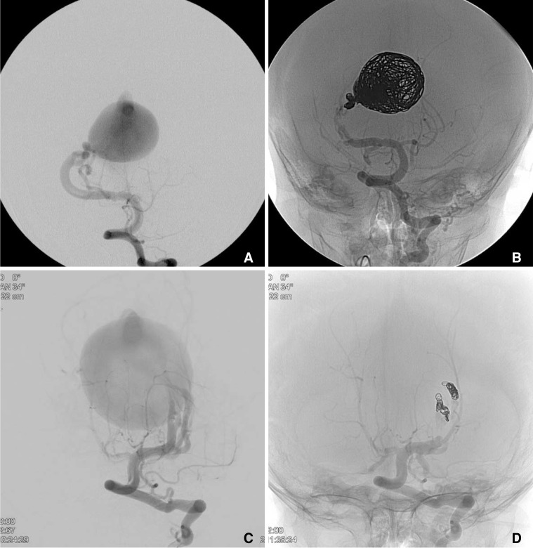
Materials and methods: Five pediatric VGAM patients (age range; 11 weeks to 2 yrs 2 mos) with high flow fistulous angioarchitecture were treated with adjuvant coils 1) in the distal feeding artery and/or 2) in the vein of Galen followed by glue embolization of the shunt. The angiographic / clinical outcomes were assessed.
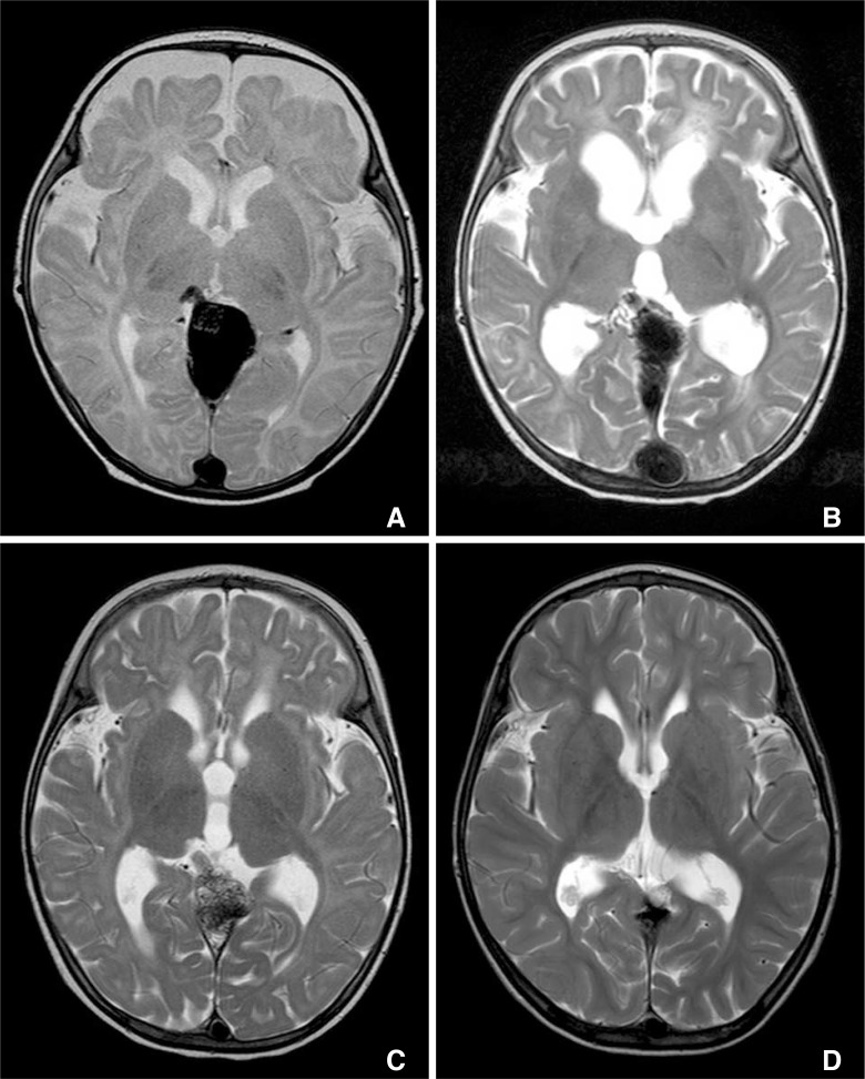
Results: Adjuvant coils were deployed in the distal feeding artery (n=3), vein of Galen pouch plus distal feeding artery (n=2). Additional transarterial glue embolization of the fistulae was successfully performed (n=4). Complete occlusion was achieved with coils in one case. Complete occlusion was achieved for all mural type cases (n=4). Residual feeders remained in a case of choroidal type of VGAM. No complications were noted related to the treatment. All patients showed normal development on follow up (range: 7.6 to 88.8 mo, mean 49.3 mo). Initial hydrocephalus improved on follow up despite coil mass effect in dilated vein of Galen.
Conclusion: Adjuvant coils for flow control with glue embolization may be a safe and effective treatment method for VGAM patients with high flow fistulous feeders.
Keywords: Coil; Embolization; Glue; Vein of Galen aneurysmal malformation.
Figures
Similar articles
-
Reversal of Fetal Compromise Following In Utero Treatment of Vein of Galen Malformation Using Glue.Prenat Diagn. 2024 Oct;44(11):1367-1371. doi: 10.1002/pd.6662. Epub 2024 Sep 7. Prenat Diagn. 2024. PMID: 39243201
-
Cognitive and functional status after vein of Galen aneurysmal malformation endovascular occlusion.World J Radiol. 2012 Mar 28;4(3):83-9. doi: 10.4329/wjr.v4.i3.83. World J Radiol. 2012. PMID: 22468188 Free PMC article.
-
Susceptibility weighted imaging in infants with staged embolization of vein of Galen aneurysmal malformations.J Neuroradiol. 2019 May;46(3):214-221. doi: 10.1016/j.neurad.2018.09.009. Epub 2018 Nov 10. J Neuroradiol. 2019. PMID: 30423378
-
The management of vein of Galen aneurysmal malformations.Neurosurgery. 2006 Nov;59(5 Suppl 3):S184-94; discussion S3-13. doi: 10.1227/01.NEU.0000237445.39514.16. Neurosurgery. 2006. PMID: 17053602 Review.
-
[Vein of Galen aneurysmal malformation].Radiologie (Heidelb). 2022 Aug;62(8):671-674. doi: 10.1007/s00117-022-01029-z. Epub 2022 Jun 23. Radiologie (Heidelb). 2022. PMID: 35736995 Review. German.
Cited by
-
Rapid Ventricular Pacing Facilitates Transarterial Embolization in Vein of Galen Malformations.Interv Neuroradiol. 2023 Apr;29(2):183-188. doi: 10.1177/15910199221082472. Epub 2022 Mar 2. Interv Neuroradiol. 2023. PMID: 35234073 Free PMC article.
-
Rare Neurovascular Diseases in Korea: Classification and Related Genetic Variants.Korean J Radiol. 2021 Aug;22(8):1379-1396. doi: 10.3348/kjr.2020.1171. Epub 2021 May 20. Korean J Radiol. 2021. PMID: 34047503 Free PMC article. Review.
-
Chemical abscess post vein of Galen aneurysmal malformation embolisation with ethylene vinyl alcohol copolymer.SA J Radiol. 2024 Apr 30;28(1):2841. doi: 10.4102/sajr.v28i1.2841. eCollection 2024. SA J Radiol. 2024. PMID: 38725970 Free PMC article.
-
Long Vascular Sheaths for Transfemoral Neuroendovascular Procedures in Children.Neurointervention. 2021 Jul;16(2):149-157. doi: 10.5469/neuroint.2021.00192. Epub 2021 Jun 3. Neurointervention. 2021. PMID: 34078026 Free PMC article.
References
-
- Lasjaunias PL, Chng SM, Sachet M, Alvarez H, Rodesch G, Garcia-Monaco R. The management of vein of galen aneurysmal malformations. Neurosurgery. 2006;59:S184–S194. discussion S3-13. - PubMed
-
- Lasjaunias P, Ter Brugge K, Berenstein A. Surgical neuroangiography: Clinical and interventional aspects in children. 2nd ed. Berlin Heidelberg: Springer-Verlag; 2006.
-
- Khullar D, Andeejani AM, Bulsara KR. Evolution of treatment options for vein of galen malformations. J Neurosurg Pediatr. 2010;6:444–451. - PubMed
-
- Berenstein A, Fifi JT, Niimi Y, Presti S, Ortiz R, Ghatan S, et al. Vein of Galen malformations in neonates: new management paradigms for improving outcomes. Neurosurgery. 2012;70:1207–1213. discussion 1213-1204. - PubMed
LinkOut - more resources
Full Text Sources
Other Literature Sources