

Review
doi: 10.7759/cureus.2035.
A Comprehensive Review of Medical Imaging Equipment Used in Cadaveric Studies
Affiliations
- PMID: 29535907
- PMCID: PMC5841925
- DOI: 10.7759/cureus.2035
Item in Clipboard
Review
A Comprehensive Review of Medical Imaging Equipment Used in Cadaveric Studies
Cureus.
.
Abstract
Medical imaging techniques have led to great advances in clinical anatomy and forensic pathology. New and emerging technologies allow healthcare professionals to view and understand the human body from different perspectives. This gives way to new and improved interventions, treatment plans, and an overall understanding of the human body. Herein, we present a comprehensive review of the various medical imaging equipment used in cadaveric studies along with their individual strengths and limitations.
Keywords: anatomy; cadaver; computed tomography; endoscopy; magnetic resonance imaging; ultrasound.
Conflict of interest statement
The authors have declared that no competing interests exist.
References
-
- Postmortem CT angiography: capabilities and limitations in traumatic and natural causes of death. Ross SG, Bolliger SA, Ampanozi G, et al. Radiographics. 2014;34:830–846. - PubMed
-
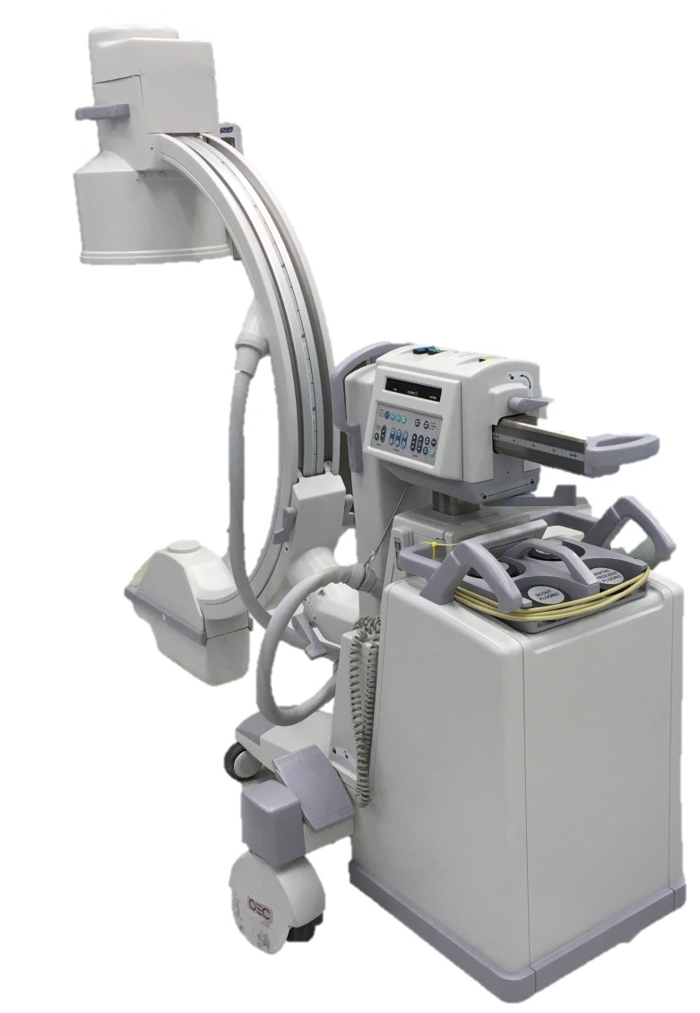
- Ultra-high resolution C-Arm CT arthrography of the wrist: Radiation dose and image quality compared to conventional multidetector computed tomography. Werncke T, Sonnow L, Meyer BC, et al. Eur J Radiol. 2017;89:191–199. - PubMed
-
- Injuries in full-scale vehicle side impact moving deformable barrier and pole tests using postmortem human subjects. Yoganandan N, Pintar F, Humm J, et al. Traffic Inj Prev. 2015;16:224–230. - PubMed
Publication types
LinkOut - more resources
Full Text Sources
Other Literature Sources