Review
. 2018 Dec;12(4):529-533.
doi: 10.1007/s12105-018-0907-5. Epub 2018 Mar 13.

Primary Leiomyosarcoma of the Buccal Mucosa: Report of a Case and Review of the Literature

Affiliations
Review

Primary Leiomyosarcoma of the Buccal Mucosa: Report of a Case and Review of the Literature

Eugene M Ko et al. Head Neck Pathol. 2018 Dec.

Abstract

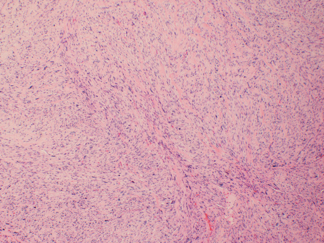
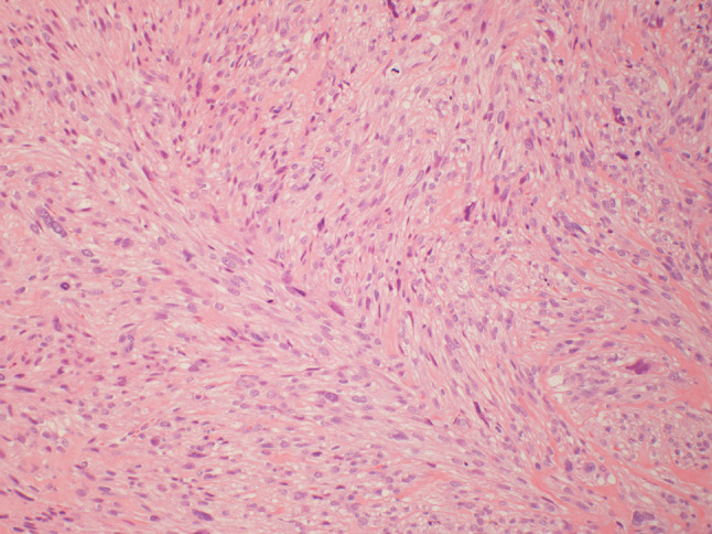
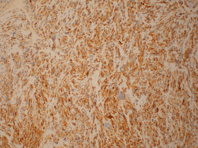
This clinicopathologic study of primary oral leiomyosarcoma of the buccal mucosa involves a literature review of 15 cases with the addition of our report of a case. The demographic details, tumor size, treatment and outcome are documented for all the cases. In addition, this review examines the histologic features of leiomyosarcoma while noting that differentiation from other spindle cell tumors can be challenging, underscoring the necessity of an immunohistochemical work up for an accurate diagnosis. The unpredictability of the clinical behavior of these aggressive tumors requires, at the very least, wide local surgical excision and prolonged follow up.

Keywords: Head and neck; Leiomyosarcoma; Oral cavity.


Conflict of interest statement

Authors Eugene M. Ko and Jonathan McHugh declares that they have no conflict of interest.

Figures

Fig. 1
Fig. 1
At low power (×40), the tumor is comprised of spindle cells arranged in long fascicles. Scattered cells with marked nuclear atypia are present
Fig. 2
Fig. 2
At medium power (×100), the spindle cells are arranged in fascicles and contain abundant fibrillary eosinophilic cytoplasm. The nuclei tend to be oval with perinuclear vacuoles. Mitotic figures are common as are scattered cells with marked nuclear atypia
Fig. 3
Fig. 3
Desmin staining (×100) demonstrates diffuse strong staining in the cytoplasm of the neoplastic cells
Fig. 4
Fig. 4
Muscle specific actin (×100) demonstrates diffuse moderate staining in the cytoplasm of the neoplastic cells
Fig. 5
Fig. 5
Smooth muscle actin (×100) demonstrates diffuse strong staining in the cytoplasm of the neoplastic cells

References

    1. Solar AA, Jordan RC. Soft tissue tumors and common metastases of the oral cavity. Periodontol 2000. 2011;57(1):177–197. doi: 10.1111/j.1600-0757.2011.00388.x. - DOI - PubMed
    1. Schutz A, Smeets R, Driemel O, Hakim SG, Kosmehl H, Hanken H, Kolk A. Primary and secondary leiomyosarcoma of the oral and perioral region–clinicopathological and immunohistochemical analysis of a rare entity with a review of the literature. J Oral Maxillofac Surg. 2013;71(6):1132–1142. doi: 10.1016/j.joms.2012.12.011. - DOI - PubMed
    1. Schenberg ME, Slootweg PJ, Koole R. Leiomyosarcomas of the oral cavity. Report of four cases and review of the literature. J Cranio-Maxillofac Surg. 1993;21(8):342–347. doi: 10.1016/S1010-5182(05)80495-0. - DOI - PubMed
    1. Yan B, Li Y, Pan J, Xia H, Li LJ. Primary oral leiomyosarcoma: a retrospective clinical analysis of 20 cases. Oral Dis. 2010;16(2):198–203. doi: 10.1111/j.1601-0825.2009.01635.x. - DOI - PubMed
    1. Lo Muzio L, Favia G, Mignogna MD, Piattelli A, Maiorano E. Primary intraoral leiomyosarcoma of the tongue: an immunohistochemical study and review of the literature. Oral Oncol. 2000;36(6):519–524. doi: 10.1016/S1368-8375(00)00044-0. - DOI - PubMed

LinkOut - more resources