

Application of the CometChip platform to assess DNA damage in field-collected blood samples from turtles
- PMID: 29536573
- PMCID: PMC5902651
- DOI: 10.1002/em.22183
Application of the CometChip platform to assess DNA damage in field-collected blood samples from turtles
Abstract
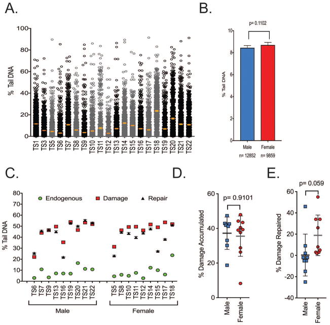
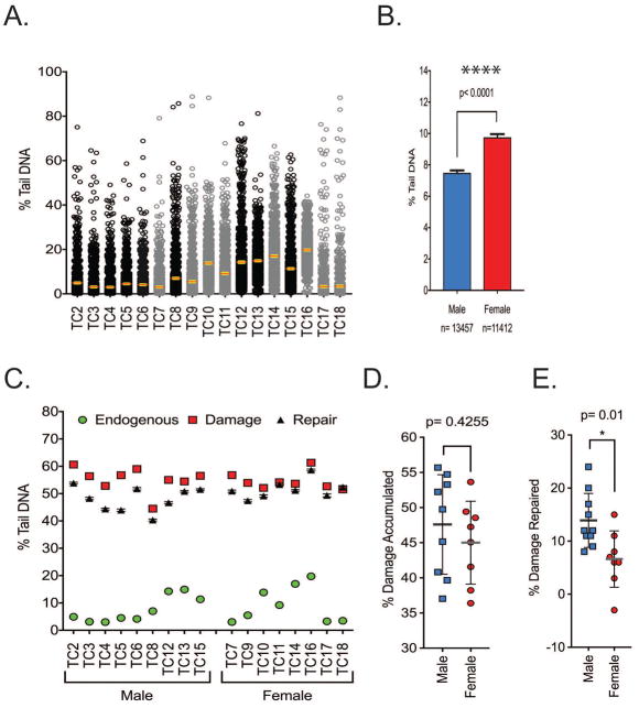
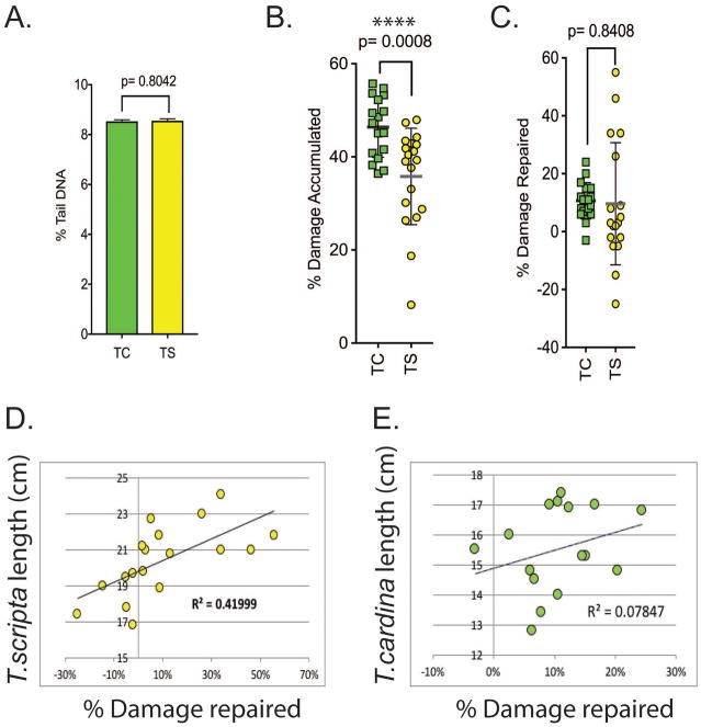
DNA damage has been linked to genomic instability and the progressive breakdown of cellular and organismal homeostasis, leading to the onset of disease and reduced longevity. Insults to DNA from endogenous sources include base deamination, base hydrolysis, base alkylation, and metabolism-induced oxidative damage that can lead to single-strand and double-strand DNA breaks. Alternatively, exposure to environmental pollutants, radiation or ultra-violet light, can also contribute to exogenously derived DNA damage. We previously validated a novel, high through-put approach to measure levels of DNA damage in cultured mammalian cells. This new CometChip Platform builds on the classical single cell gel electrophoresis or comet methodology used extensively in environmental toxicology and molecular biology. We asked whether the CometChip Platform could be used to measure DNA damage in samples derived from environmental field studies. To this end, we determined that nucleated erythrocytes from multiple species of turtle could be successfully evaluated in the CometChip Platform to quantify levels of DNA damage. In total, we compared levels of DNA damage in 40 animals from two species: the box turtle (Terrapene carolina) and the red-eared slider (Trachemys scripta elegans). Endogenous levels of DNA damage were identical between the two species, yet we did discover some sex-linked differences and changes in DNA damage accumulation. Based on these results, we confirm that the CometChip Platform allows for the measurement of DNA damage in a large number of samples quickly and accurately, and is particularly adaptable to environmental studies using field-collected samples. Environ. Mol. Mutagen. 59:322-333, 2018. © 2018 Wiley Periodicals, Inc.
Keywords: CometChip; DNA damage; comet assay; erythrocytes; vertebrates.
© 2018 Wiley Periodicals, Inc.
Figures
Similar articles
-
Application of the micronucleus test and comet assay in Trachemys callirostris erythrocytes as a model for in situ genotoxic monitoring.Ecotoxicol Environ Saf. 2016 May;127:108-16. doi: 10.1016/j.ecoenv.2016.01.016. Epub 2016 Jan 22. Ecotoxicol Environ Saf. 2016. PMID: 26809080
-
The development and validation of EpiComet-Chip, a modified high-throughput comet assay for the assessment of DNA methylation status.Environ Mol Mutagen. 2017 Aug;58(7):508-521. doi: 10.1002/em.22101. Epub 2017 Jul 29. Environ Mol Mutagen. 2017. PMID: 28755435 Free PMC article.
-
Applications of CometChip for Environmental Health Studies.Chem Res Toxicol. 2020 Jul 20;33(7):1528-1538. doi: 10.1021/acs.chemrestox.9b00393. Epub 2020 Jun 10. Chem Res Toxicol. 2020. PMID: 32519858 Free PMC article. Review.
-
Genotoxicity of environmental agents assessed by the alkaline comet assay.Basic Clin Pharmacol Toxicol. 2005;96 Suppl 1:1-42. Basic Clin Pharmacol Toxicol. 2005. PMID: 15859009 Review.
-
Assessing CometChip technology for DNA damage studies in non-model species: distinct UV-induced responses in turtles and mammals.BMC Res Notes. 2025 Jun 2;18(1):243. doi: 10.1186/s13104-025-07285-1. BMC Res Notes. 2025. PMID: 40457500 Free PMC article.
Cited by
-
Utilization of the CometChip assay for detecting PAH-induced DNA bulky adducts in a 3D primary human bronchial epithelial cell model.Toxicology. 2025 Nov;517:154241. doi: 10.1016/j.tox.2025.154241. Epub 2025 Jul 23. Toxicology. 2025. PMID: 40713011 Free PMC article.
-
Identifying Compounds with Genotoxicity Potential Using Tox21 High-Throughput Screening Assays.Chem Res Toxicol. 2019 Jul 15;32(7):1384-1401. doi: 10.1021/acs.chemrestox.9b00053. Epub 2019 Jun 18. Chem Res Toxicol. 2019. PMID: 31243984 Free PMC article.
-
Reduced DNA Repair Capacity in Prostate Cancer Patients: A Phenotypic Approach Using the CometChip.Cancers (Basel). 2022 Jun 25;14(13):3117. doi: 10.3390/cancers14133117. Cancers (Basel). 2022. PMID: 35804887 Free PMC article.
-
Urothelial genotoxicity of environmental chemicals detected in the urine of healthy dogs and their owners.J Clin Transl Sci. 2024 Oct 24;8(1):e172. doi: 10.1017/cts.2024.546. eCollection 2024. J Clin Transl Sci. 2024. PMID: 39619067 Free PMC article.
-
DNA Repair Capacity and Clinicopathological Characteristics in Puerto Rican Hispanic/Latino Patients with Metastatic Castration-Resistant Prostate Cancer.Cancers (Basel). 2025 Jan 16;17(2):279. doi: 10.3390/cancers17020279. Cancers (Basel). 2025. PMID: 39858060 Free PMC article.
References
-
- Azqueta A, Collins AR. The essential comet assay: a comprehensive guide to measuring DNA damage and repair. Arch Toxicol. 2013;87(6):949–968. - PubMed
-
- Belpaeme K, Cooreman K, Kirsch-Volders M. Development and validation of the in vivo alkaline comet assay for detecting genomic damage in marine flatfish. Mutat Res. 1998;415(3):167–184. - PubMed
-
- Bhagat J. Combinations of genotoxic tests for the evaluation of group 1 IARC carcinogens. J Appl Toxicol. 2018;38(1):81–99. - PubMed
-
- Blanpain C, Mohrin M, Sotiropoulou PA, Passegue E. DNA-damage response in tissue-specific and cancer stem cells. Cell Stem Cell. 2011;8(1):16–29. - PubMed
-
- Bonisoli-Alquati A, Voris A, Mousseau TA, Moller AP, Saino N, Wyatt MD. DNA damage in barn swallows (Hirundo rustica) from the Chernobyl region detected by use of the comet assay. Comp Biochem Physiol C Toxicol Pharmacol. 2010;151(3):271–277. - PubMed
Publication types
MeSH terms
Substances
Grants and funding
LinkOut - more resources
Full Text Sources
Other Literature Sources
