

Lightweight, compact, and high-performance 3T MR system for imaging the brain and extremities
- PMID: 29536587
- PMCID: PMC6107412
- DOI: 10.1002/mrm.27175
Lightweight, compact, and high-performance 3T MR system for imaging the brain and extremities
Abstract
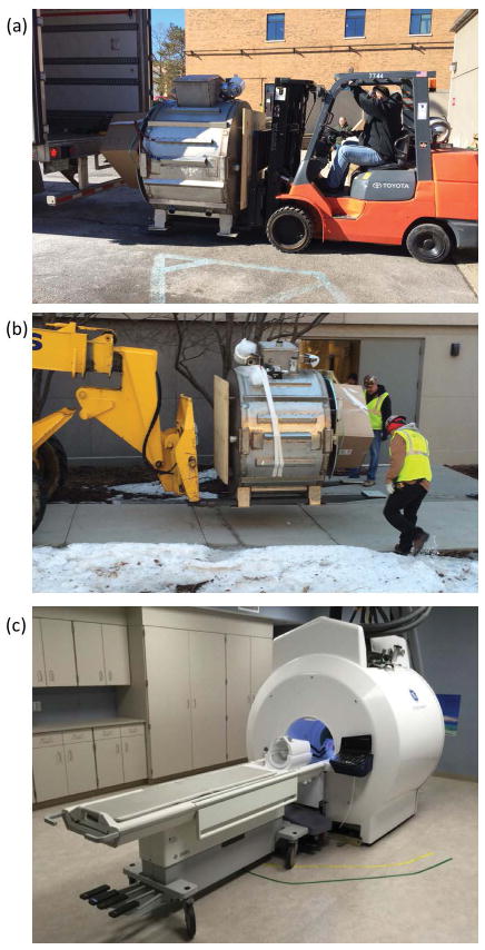
Purpose: To build and evaluate a small-footprint, lightweight, high-performance 3T MRI scanner for advanced brain imaging with image quality that is equal to or better than conventional whole-body clinical 3T MRI scanners, while achieving substantial reductions in installation costs.
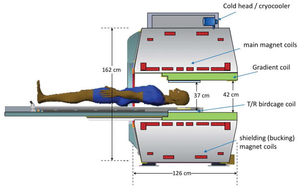
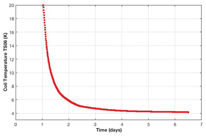
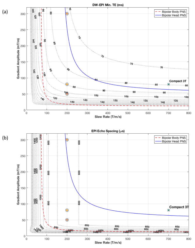
Methods: A conduction-cooled magnet was developed that uses less than 12 liters of liquid helium in a gas-charged sealed system, and standard NbTi wire, and weighs approximately 2000 kg. A 42-cm inner-diameter gradient coil with asymmetric transverse axes was developed to provide patient access for head and extremity exams, while minimizing magnet-gradient interactions that adversely affect image quality. The gradient coil was designed to achieve simultaneous operation of 80-mT/m peak gradient amplitude at a slew rate of 700 T/m/s on each gradient axis using readily available 1-MVA gradient drivers.
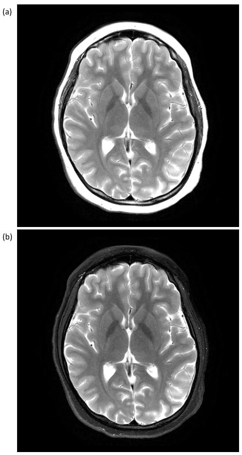
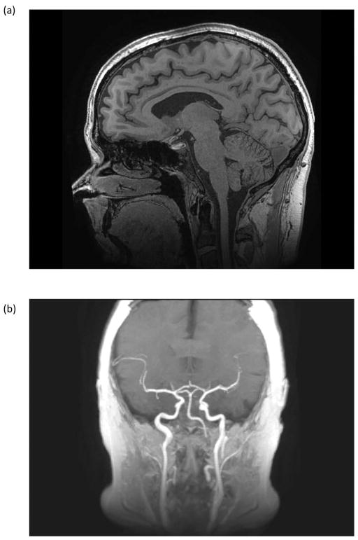
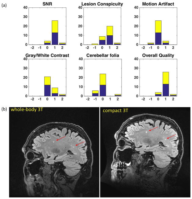
Results: In a comparison of anatomical imaging in 16 patients using T2 -weighted 3D fluid-attenuated inversion recovery (FLAIR) between the compact 3T and whole-body 3T, image quality was assessed as equivalent to or better across several metrics. The ability to fully use a high slew rate of 700 T/m/s simultaneously with 80-mT/m maximum gradient amplitude resulted in improvements in image quality across EPI, DWI, and anatomical imaging of the brain.
Conclusions: The compact 3T MRI system has been in continuous operation at the Mayo Clinic since March 2016. To date, over 200 patient studies have been completed, including 96 comparison studies with a clinical 3T whole-body MRI. The increased gradient performance has reliably resulted in consistently improved image quality.
© 2018 International Society for Magnetic Resonance in Medicine.
Figures
Similar articles
-
Peripheral nerve stimulation limits of a high amplitude and slew rate magnetic field gradient coil for neuroimaging.Magn Reson Med. 2020 Jan;83(1):352-366. doi: 10.1002/mrm.27909. Epub 2019 Aug 6. Magn Reson Med. 2020. PMID: 31385628 Free PMC article.
-
Reduced acoustic noise in diffusion tensor imaging on a compact MRI system.Magn Reson Med. 2018 Jun;79(6):2902-2911. doi: 10.1002/mrm.26949. Epub 2017 Oct 2. Magn Reson Med. 2018. PMID: 28971512 Free PMC article.
-
Highly efficient head-only magnetic field insert gradient coil for achieving simultaneous high gradient amplitude and slew rate at 3.0T (MAGNUS) for brain microstructure imaging.Magn Reson Med. 2020 Jun;83(6):2356-2369. doi: 10.1002/mrm.28087. Epub 2019 Nov 25. Magn Reson Med. 2020. PMID: 31763726
-
[3 Tesla MRI: successful results with higher field strengths].Radiologe. 2004 Jan;44(1):31-47. doi: 10.1007/s00117-003-1000-x. Radiologe. 2004. PMID: 14997868 Review. German.
-
Connectome 2.0: Developing the next-generation ultra-high gradient strength human MRI scanner for bridging studies of the micro-, meso- and macro-connectome.Neuroimage. 2021 Nov;243:118530. doi: 10.1016/j.neuroimage.2021.118530. Epub 2021 Aug 28. Neuroimage. 2021. PMID: 34464739 Free PMC article. Review.
Cited by
-
MRI Insights Into Adolescent Neurocircuitry-A Vision for the Future.Front Hum Neurosci. 2020 Jul 7;14:237. doi: 10.3389/fnhum.2020.00237. eCollection 2020. Front Hum Neurosci. 2020. PMID: 32733218 Free PMC article.
-
Evaluation of a compact 3 T MRI scanner for patients with implanted devices.Magn Reson Imaging. 2023 Nov;103:109-118. doi: 10.1016/j.mri.2023.07.009. Epub 2023 Jul 17. Magn Reson Imaging. 2023. PMID: 37468020 Free PMC article.
-
Prediction of Surgical Outcomes in Normal Pressure Hydrocephalus by MR Elastography.AJNR Am J Neuroradiol. 2024 Mar 7;45(3):328-334. doi: 10.3174/ajnr.A8108. AJNR Am J Neuroradiol. 2024. PMID: 38272572 Free PMC article.
-
Time-dependent diffusion MRI probes cerebellar microstructural alterations in a mouse model of Down syndrome.Brain Commun. 2021 Apr 5;3(2):fcab062. doi: 10.1093/braincomms/fcab062. eCollection 2021. Brain Commun. 2021. PMID: 33937769 Free PMC article.
-
The effect of spiral trajectory correction on pseudo-continuous arterial spin labeling with high-performance gradients on a compact 3T scanner.Magn Reson Med. 2020 Jul;84(1):192-205. doi: 10.1002/mrm.28110. Epub 2019 Dec 4. Magn Reson Med. 2020. PMID: 31799747 Free PMC article.
References
-
- Turner R. Gradient coil design: a review of methods. Magn Reson Imaging. 1993;11:903–920. - PubMed
-
- Reilly JP. Maximum pulsed electromagnetic field limits based on peripheral nerve stimulation: application to IEEE/ANSI C95.1 electromagnetic field standards. IEEE Trans Biomed Eng. 1998;45:137–141. - PubMed
-
- Reilly JP. Peripheral nerve stimulation by induced electric currents: exposure to time-varying magnetic fields. Med Biol Eng Comput. 1989;27:101–110. - PubMed
-
- Chronik BA, Rutt BK. A comparison between human magnetostimulation thresholds in whole-body and head/neck gradient coils. Magn Reson Med. 2001;46:386–394. - PubMed
Publication types
MeSH terms
Grants and funding
LinkOut - more resources
Full Text Sources
Other Literature Sources
Medical
