

Differences in proliferation rate between CADASIL and control vascular smooth muscle cells are related to increased TGFβ expression
- PMID: 29536621
- PMCID: PMC5980144
- DOI: 10.1111/jcmm.13534
Differences in proliferation rate between CADASIL and control vascular smooth muscle cells are related to increased TGFβ expression
Abstract
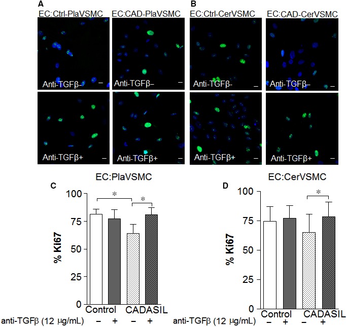
Cerebral autosomal-dominant arteriopathy with subcortical infarcts and leukoencephalopathy (CADASIL) is a familial fatal progressive degenerative disorder. One of the pathological hallmarks of CADASIL is a dramatic reduction of vascular smooth muscle cells (VSMCs) in cerebral arteries. Using VSMCs from the vasculature of the human umbilical cord, placenta and cerebrum of CADASIL patients, we found that CADASIL VSMCs had a lower proliferation rate compared to control VSMCs. Exposure of control VSMCs and endothelial cells (ECs) to media derived from CADASIL VSMCs lowered the proliferation rate of all cells examined. By quantitative RT-PCR analysis, we observed increased Transforming growth factor-β (TGFβ) gene expression in CADASIL VSMCs. Adding TGFβ-neutralizing antibody restored the proliferation rate of CADASIL VSMCs. We assessed proliferation differences in the presence or absence of TGFβ-neutralizing antibody in ECs co-cultured with VSMCs. ECs co-cultured with CADASIL VSMCs exhibited a lower proliferation rate than those co-cultured with control VSMCs, and neutralization of TGFβ normalized the proliferation rate of ECs co-cultured with CADASIL VSMCs. We suggest that increased TGFβ expression in CADASIL VSMCs is involved in the reduced VSMC proliferation in CADASIL and may play a role in situ in altered proliferation of neighbouring cells in the vasculature.
Keywords: CADASIL; NOTCH3; Transforming growth factor-β; endothelial cells; vascular smooth muscle cells.
© 2018 The Authors. Journal of Cellular and Molecular Medicine published by John Wiley & Sons Ltd and Foundation for Cellular and Molecular Medicine.
Figures


Similar articles
-
Notch3 in Development, Health and Disease.Biomolecules. 2020 Mar 23;10(3):485. doi: 10.3390/biom10030485. Biomolecules. 2020. PMID: 32210034 Free PMC article. Review.
-
CADASIL mutations and shRNA silencing of NOTCH3 affect actin organization in cultured vascular smooth muscle cells.J Cereb Blood Flow Metab. 2012 Dec;32(12):2171-80. doi: 10.1038/jcbfm.2012.123. Epub 2012 Sep 5. J Cereb Blood Flow Metab. 2012. PMID: 22948298 Free PMC article. Clinical Trial.
-
Experimental studies of mitochondrial function in CADASIL vascular smooth muscle cells.Exp Cell Res. 2013 Feb 1;319(3):134-43. doi: 10.1016/j.yexcr.2012.09.015. Epub 2012 Oct 1. Exp Cell Res. 2013. PMID: 23036509
-
Nuclear abnormalities in vascular myocytes in cerebral autosomal-dominant arteriopathy with subcortical infarcts and leukoencephalopathy (CADASIL).Neuropathology. 2018 Dec;38(6):601-608. doi: 10.1111/neup.12519. Epub 2018 Nov 6. Neuropathology. 2018. PMID: 30402942
-
An overview of Notch3 function in vascular smooth muscle cells.Prog Biophys Mol Biol. 2008 Jan-Apr;96(1-3):499-509. doi: 10.1016/j.pbiomolbio.2007.07.006. Epub 2007 Jul 29. Prog Biophys Mol Biol. 2008. PMID: 17854869 Review.
Cited by
-
Update on the Epidemiology, Pathogenesis, and Biomarkers of Cerebral Autosomal Dominant Arteriopathy With Subcortical Infarcts and Leukoencephalopathy.J Clin Neurol. 2023 Jan;19(1):12-27. doi: 10.3988/jcn.2023.19.1.12. J Clin Neurol. 2023. PMID: 36606642 Free PMC article. Review.
-
Signaling pathways and molecular mechanisms involved in the onset and progression of cerebral autosomal dominant arteriopathy with subcortical infarcts and leukoencephalopathy (CADASIL); a focus on Notch3 signaling.J Headache Pain. 2025 Apr 29;26(1):96. doi: 10.1186/s10194-025-02025-z. J Headache Pain. 2025. PMID: 40301727 Free PMC article. Review.
-
Modeling CADASIL vascular pathologies with patient-derived induced pluripotent stem cells.Protein Cell. 2019 Apr;10(4):249-271. doi: 10.1007/s13238-019-0608-1. Epub 2019 Feb 18. Protein Cell. 2019. PMID: 30778920 Free PMC article.
-
Notch3 in Development, Health and Disease.Biomolecules. 2020 Mar 23;10(3):485. doi: 10.3390/biom10030485. Biomolecules. 2020. PMID: 32210034 Free PMC article. Review.
-
ER stress induced immunopathology involving complement in CADASIL: implications for therapeutics.Acta Neuropathol Commun. 2023 May 8;11(1):76. doi: 10.1186/s40478-023-01558-1. Acta Neuropathol Commun. 2023. PMID: 37158955 Free PMC article.
References
-
- Chabriat H, Joutel A, Dichgans M, Tournier‐Lasserve E, Bousser MG. Cadasil. Lancet Neurol. 2009;8:643‐653. - PubMed
-
- Joutel A, Vahedi K, Corpechot C, et al. Strong clustering and stereotyped nature of Notch3 mutations in CADASIL patients. Lancet. 1997;350:1511‐1515. - PubMed
-
- Chabriat H, Levy C, Taillia H, et al. Patterns of MRI lesions in CADASIL. Neurology. 1998;51:452‐457. - PubMed
-
- Chabriat H, Vahedi K, Iba‐Zizen MT, et al. Clinical spectrum of CADASIL: a study of 7 families. cerebral autosomal dominant arteriopathy with subcortical infarcts and leukoencephalopathy. Lancet. 1995;346:934‐939. - PubMed
-
- Desmond DW, Moroney JT, Lynch T, Chan S, Chin SS, Mohr JP. The natural history of CADASIL: a pooled analysis of previously published cases. Stroke. 1999;30:1230‐1233. - PubMed
Publication types
MeSH terms
Substances
LinkOut - more resources
Full Text Sources
Other Literature Sources
Miscellaneous
