Clinical and imaging features of Kaposiform Hemangioendothelioma
- PMID: 29536769
- PMCID: PMC6223283
- DOI: 10.1259/bjr.20170798
Clinical and imaging features of Kaposiform Hemangioendothelioma
Abstract
Objective: Kaposiform hemangioendothelioma (KHE) is a unique locally aggressive vascular tumor with poor prognosis. The aim of this study is to assess the clinical and imaging features of KHE, and to compare the differences between solitary and diffusive infiltrative subtype further.
Methods: The clinical and radiological findings of a cohort of 25 cases with histologically proven KHE, between June 2011 and June 2016, were reviewed retrospectively. 7 solitary and 18 diffusive infiltrative subtypes KHE were included. The differences of clinical and imaging features between these two subtypes were compared statistically by Wilcoxon rank sum test and Fisher exact test.
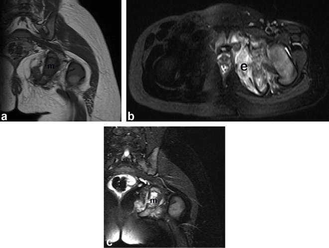
Results: The median age was 4 months old. 20 cases (80%) were accompanied by Kasabach-Merritt phenomenon (KMP). Most KHE located in trunk and/or extremity. The masses showed inhomogeneous echogenicity and were rich in vascularity on ultrasound; showed isoattenuation relative to muscle on unenhanced CT, isointense (n = 15) or slightly hyperintense (n = 7) T1 weighted imaging (T1WI) signal relative to muscle, mainly heterogeneous hyperintense or slightly hyperintense with speckled hypointense (n = 17) T2WI signal (77%) relative to muscle, and notable (n = 15) and moderate (n = 3) enhancement. Feeding and draining vessels were revealed in 15 cases. Five masses with DWI showed slightly restricted diffusivity, with average apparent diffusion coefficient value of (1.28 ± 0.09) × 10-3 mm2 s-1. Necrosis and hemorrhage were also found. Compared with solitary ones, diffusive infiltrative KHE were larger, more commonly accompanied by KMP and reticular lymphedema, and more frequently located in trunk and/or extremity.
Conclusion: Five masses with DWI showed slightly restricted diffusivity. A hypervascular mass accompanied by KMP and reticular lymphedema, with speckled hypointense signal T2WI signal, especially in pediatric patients, is highly suggestive of the diagnosis of KHE. Advances in knowledge: Speckled hypointense signal T2WI signal, and notable enhancement were unique features of KHE. KHE showed slightly restricted diffusivity on DWI, commonly accompanied by KMP and reticular lymphedema.
Figures


Similar articles
-
Ultrasonography and magnetic resonance imaging features of kaposiform hemangioendothelioma and tufted angioma.J Dermatol. 2019 Oct;46(10):835-842. doi: 10.1111/1346-8138.15025. Epub 2019 Aug 2. J Dermatol. 2019. PMID: 31373042
-
Imaging findings of Kaposiform Hemangioendothelioma in children.Eur J Radiol. 2017 Jan;86:198-205. doi: 10.1016/j.ejrad.2016.11.015. Epub 2016 Nov 10. Eur J Radiol. 2017. PMID: 28027747
-
Atypically presenting kaposiform hemangioendothelioma of the knee: ultrasound findings.J Med Ultrason (2001). 2018 Oct;45(4):653-656. doi: 10.1007/s10396-018-0878-x. Epub 2018 Apr 10. J Med Ultrason (2001). 2018. PMID: 29637402
-
Kaposiform hemangioendothelioma in children: a benign vascular tumor with multiple treatment options.World J Pediatr. 2018 Aug;14(4):322-329. doi: 10.1007/s12519-018-0171-5. Epub 2018 Jul 27. World J Pediatr. 2018. PMID: 30054848 Review.
-
Kaposiform haemangioendothelioma: a review with emphasis on histological differential diagnosis.Pathology. 2017 Jun;49(4):356-362. doi: 10.1016/j.pathol.2017.03.001. Epub 2017 Apr 21. Pathology. 2017. PMID: 28438388 Review.
Cited by
-
Kaposiform hemangioendothelioma without cutaneous involvement.J Cancer Res Clin Oncol. 2018 Dec;144(12):2475-2484. doi: 10.1007/s00432-018-2759-5. Epub 2018 Oct 6. J Cancer Res Clin Oncol. 2018. PMID: 30293120 Free PMC article.
-
Kaposiform hemangioendothelioma of the thigh: A case report.Radiol Case Rep. 2023 Jun 26;18(9):3152-3156. doi: 10.1016/j.radcr.2023.05.052. eCollection 2023 Sep. Radiol Case Rep. 2023. PMID: 37409100 Free PMC article.
-
Digital subtraction angiography and trans arterial embolization in preventing massive hemorrhage of Kaposiform hemangioendothelioma: A case report.Radiol Case Rep. 2022 Sep 17;17(11):4392-4398. doi: 10.1016/j.radcr.2022.08.062. eCollection 2022 Nov. Radiol Case Rep. 2022. PMID: 36188091 Free PMC article.
-
Advancing Precision Medicine: The Role of Genetic Testing and Sequencing Technologies in Identifying Biological Markers for Rare Cancers.Cancer Med. 2025 Apr;14(8):e70853. doi: 10.1002/cam4.70853. Cancer Med. 2025. PMID: 40249565 Free PMC article. Review.
-
Kaposiform Hemangioendothelioma in Adolescent-Onset Scoliosis: A Case Report and Review of Literature.Case Rep Orthop. 2020 Feb 7;2020:1839053. doi: 10.1155/2020/1839053. eCollection 2020. Case Rep Orthop. 2020. PMID: 32089925 Free PMC article.
References
-
- International Society for the Study of Vascular Anomalies. ISSVA classification of vascular anomalies ©2014. 2017. Available from: issva.org/classification [Dec 20, 2017].
-
- Iacobas I, Simon ML, Amir T, Gribbin CE, McPartland TG, Kaufman MR, et al. . Decreased vascularization of retroperitoneal kaposiform hemangioendothelioma induced by treatment with sirolimus explains relief of symptoms. Clin Imaging 2015; 39: 529–32. doi: 10.1016/j.clinimag.2015.01.003 - DOI - PubMed
MeSH terms
Supplementary concepts
LinkOut - more resources
Full Text Sources
Other Literature Sources
Medical