

doi: 10.1039/c7cc09829d.
Melting proteins confined in nanodroplets with 10.6 μm light provides clues about early steps of denaturation
Affiliations
- PMID: 29536995
- PMCID: PMC5871606
- DOI: 10.1039/c7cc09829d
Item in Clipboard
Melting proteins confined in nanodroplets with 10.6 μm light provides clues about early steps of denaturation
Chem Commun (Camb).
.
Abstract
Ubiquitin confined within nanodroplets was irradiated with a variable-power CO2 laser. Mass spectrometry analysis shows evidence for a protein "melting"-like transition within droplets prior to solvent evaporation and ion formation. Ion mobility spectrometry reveals that structures associated with early steps of denaturation are trapped because of short droplet lifetimes.
Figures
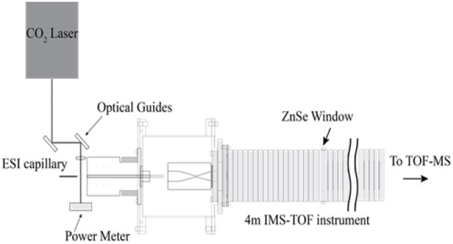
Schematic diagram of the instrument. Droplets diameters are estimated to be ~0.05 and 1.0 μm when produced from small ~1.0 and 20 μm dia. ESI capillary emitters (see Experimental section in the Supplementary Information). For 10 μM ubiquitin solutions, we estimate that only one in three of the droplets contains a protein molecule. After their formation by electrospray, droplets pass through a CO2 laser beam focused at the immediate entrance to the instrument orifice. Activation in this region may induce structural changes in the protein which leads to changes in the protein charge state distribution and ion structures. This instrument is also equipped with a ZnSe window in the middle of the drift tube, which allows ions of a known mobility to be excited with 10.6 μm radiation. A series of control experiments in which the laser is focused through the drift tube shows that gaseous protein ions are not activated in the absence of solvent at the laser powers used (See Supplementary Information). This approach has similarities with an elegant “laser spray” technique, in which 10.6 μm light from an IR laser was focused into the metal-capillary tip of an ESI source to heat the bulk solution inside the capillary (see ref. for details).

(top) Average charge state as a function of solution temperature (squares) (from ref. 9) and laser power (open circles) for ubiquitin in aqueous solution at pH 3. Solid lines show the best fit of the data assuming a two-state model with Tm = 71 ± 2 °C and Tp =10.4 ± 0.3 W. Insets show representative mass spectra at different solution temperatures and laser powers. Upon blocking the radiation, a melted charge state distribution immediately returns to the room temperature distribution, indicating that laser excitation heats only the droplets. The bottom plot shows ubiquitin ions heated in droplets at pH 2.5 (upside down triangles), pH 3.0 (open circles), and pH 4.0 (diamonds) with melting transitions of Tp = 5.8 ± 0.3, 10.4 ± 0.3, and 11.8 ± 0.3 W, respectively.
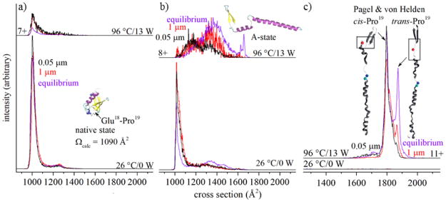
Cross section distributions for (a) [M+7H]7+, (b) [M+8H]8+, and (c) [M+11H]11+ ions of ubiquitin at different temperatures (purple) and laser powers, with black and red lines corresponding to structures that form upon irradiating droplets formed from ~1 μm and ~20 μm electrospray emitters, respectively. Structures in (c) are adapted from reference .
Similar articles
-
Understanding the Thermal Denaturation of Myoglobin with IMS-MS: Evidence for Multiple Stable Structures and Trapped Pre-equilibrium States.J Am Soc Mass Spectrom. 2021 Jan 6;32(1):64-72. doi: 10.1021/jasms.0c00075. Epub 2020 Jul 7. J Am Soc Mass Spectrom. 2021. PMID: 32539412 Free PMC article.
-
On the structural denaturation of biological analytes in trapped ion mobility spectrometry - mass spectrometry.Analyst. 2016 Jun 7;141(12):3722-30. doi: 10.1039/c5an02399h. Analyst. 2016. PMID: 26998732
-
Melting Proteins: Evidence for Multiple Stable Structures upon Thermal Denaturation of Native Ubiquitin from Ion Mobility Spectrometry-Mass Spectrometry Measurements.J Am Chem Soc. 2017 May 10;139(18):6306-6309. doi: 10.1021/jacs.7b02774. Epub 2017 Apr 26. J Am Chem Soc. 2017. PMID: 28427262
-
Confinement Dynamics of Nanodroplets between Two Surfaces: Effects of Wettability and Electric Field.Chemphyschem. 2022 Dec 16;23(24):e202200184. doi: 10.1002/cphc.202200184. Epub 2022 Sep 12. Chemphyschem. 2022. PMID: 35986551
-
Charging and supercharging of proteins for mass spectrometry: recent insights into the mechanisms of electrospray ionization.Analyst. 2019 Nov 7;144(21):6157-6171. doi: 10.1039/c9an01201j. Epub 2019 Sep 27. Analyst. 2019. PMID: 31560020 Review.
Cited by
-
Substance P in the Gas Phase: Conformational Changes and Dissociations Induced by Collisional Activation in a Drift Tube.J Am Soc Mass Spectrom. 2019 Jun;30(6):932-945. doi: 10.1007/s13361-019-02160-3. Epub 2019 Apr 12. J Am Soc Mass Spectrom. 2019. PMID: 30980379 Free PMC article.
-
Variable-Temperature Native Mass Spectrometry for Studies of Protein Folding, Stabilities, Assembly, and Molecular Interactions.Annu Rev Biophys. 2022 May 9;51:63-77. doi: 10.1146/annurev-biophys-102221-101121. Epub 2021 Dec 21. Annu Rev Biophys. 2022. PMID: 34932911 Free PMC article. Review.
-
Understanding the Thermal Denaturation of Myoglobin with IMS-MS: Evidence for Multiple Stable Structures and Trapped Pre-equilibrium States.J Am Soc Mass Spectrom. 2021 Jan 6;32(1):64-72. doi: 10.1021/jasms.0c00075. Epub 2020 Jul 7. J Am Soc Mass Spectrom. 2021. PMID: 32539412 Free PMC article.
-
Time-resolved universal temperature measurements using NaYF4:Er3+,Yb3+ upconverting nanoparticles in an electrospray jet.Beilstein J Nanotechnol. 2018 Nov 21;9:2916-2924. doi: 10.3762/bjnano.9.270. eCollection 2018. Beilstein J Nanotechnol. 2018. PMID: 30546988 Free PMC article.
-
Dissociation of Macromolecules in Laser-Heated Droplets Monitored by CD-MS.Anal Chem. 2025 Jan 21;97(2):1419-1425. doi: 10.1021/acs.analchem.4c06038. Epub 2025 Jan 7. Anal Chem. 2025. PMID: 39772511
References
-
- Lee SW, Freivogel P, Schindler T, Beauchamp J. J Am Chem Soc. 1998;120:11758.
- Silveira JA, Fort KL, Pierson NA, Clemmer DE, Russell DH. J Am Chem Soc. 2013;135:19147. - PubMed
-
- Lumry R, Eyring H. J Phys Chem. 1954;58:110.
-
- Rodrigues RC, Ortiz C, Berenguer-Murcia A, Torres R, Fernandez-Lafuente R. Chem Soc Rev. 2013;42:6290. - PubMed
- Sanchez A, Cruz J, Rueda N, dos Santos JCS, Torres R, Ortiz C, Villalonga R, Fernández-Lafuente R. RSC Adv. 2016;6:27329.
-
- Lenkinski RE, Chen DM, Glickson JD, Goldstein G. Biochim Biophys Acta. 1977;494:126. - PubMed
- Cary PD, King DS, Crane-Robinson C, Bradbury WM, Rabbani A, Goodwin GH, Johns EW. Eur J Biochem. 1980;112:577. - PubMed
- Vijay-Kumar S, Bugg CE, Cook WJ. J Mol Biol. 1987;194:531. - PubMed
- Wintrode PL, Makhatadze GI, Privalov PL. Proteins: Struct, Funct, Genet. 1994;18:246. - PubMed
- Briggs MS, Roder H. Proc Natl Acad Sci U S A. 1992;89:2017. - PMC - PubMed
- Jackson SE. Org Biomol Chem. 2006;4:1845. - PubMed
- Kony DB, Hunenberger PH, van Gunsteren WF. Protein Sci. 2007;16:1101. - PMC - PubMed
-
- Wilkinson KD, Mayer AN. Arch Biochem Biophys. 1986;250:390. - PubMed
- Harding MM, Williams DH, Woolfson DN. Biochemistry. 1991;30:3120. - PubMed
- Pan Y, Briggs MS. Biochemistry. 1992;31:11405. - PubMed
- Stockman BJ, Euvrard A, Scahill TA. J Biomol NMR. 1993;3:285. - PubMed
- Cox JPL, Evans PA, Packman LC, Williams DH, Woolfson DN. J Mol Biol. 1993;234:483. - PubMed
- Brutscher B, Brüschweiler R, Ernst RR. Biochemistry. 1997;36:13043. - PubMed
Grants and funding
LinkOut - more resources
Full Text Sources
Other Literature Sources