Comparative Study
. 2018 Jul;91(1088):20180093.
doi: 10.1259/bjr.20180093. Epub 2018 Mar 28.

Necrotizing fasciitis of the lower extremity: imaging pearls and pitfalls

Affiliations
Comparative Study

Necrotizing fasciitis of the lower extremity: imaging pearls and pitfalls

David K Tso et al. Br J Radiol. 2018 Jul.

Abstract

Objective: The purpose of this article is to review the imaging findings of necrotizing fasciitis as seen on radiograph, ultrasound, CT, and MRI, and to recognize the early findings in this potentially fatal disease.

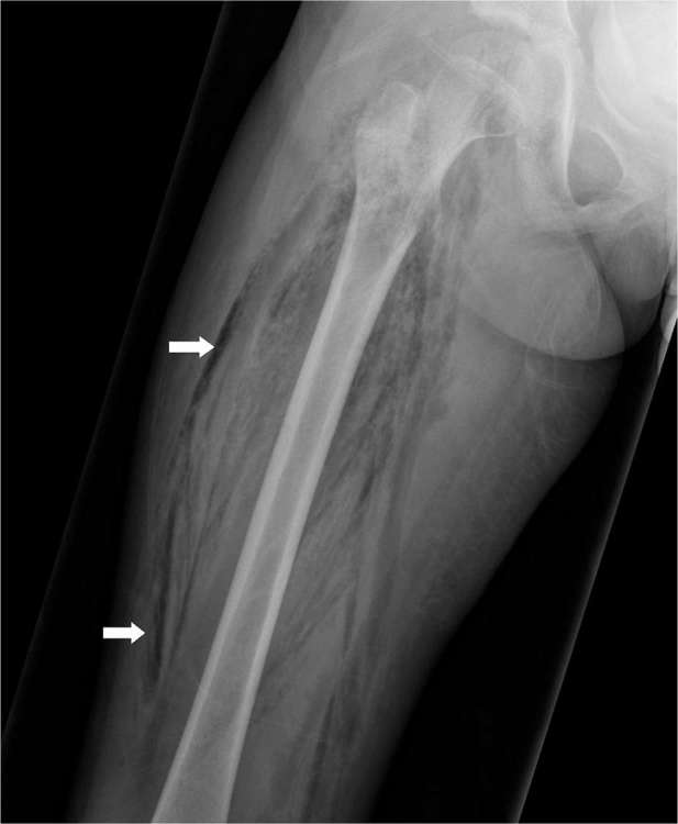
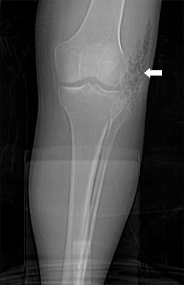
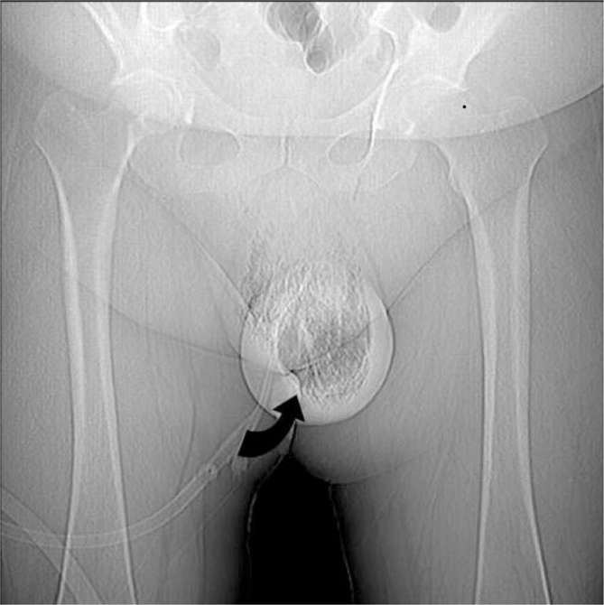
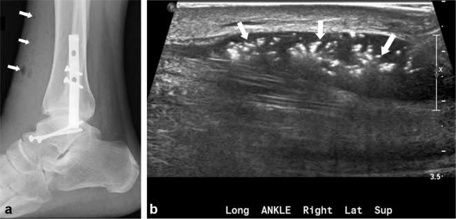
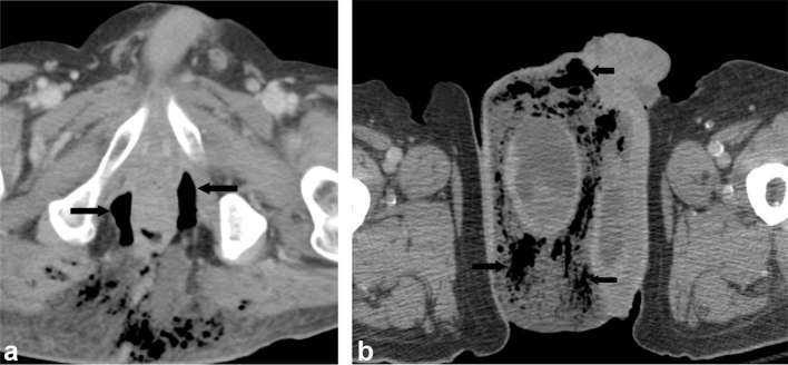
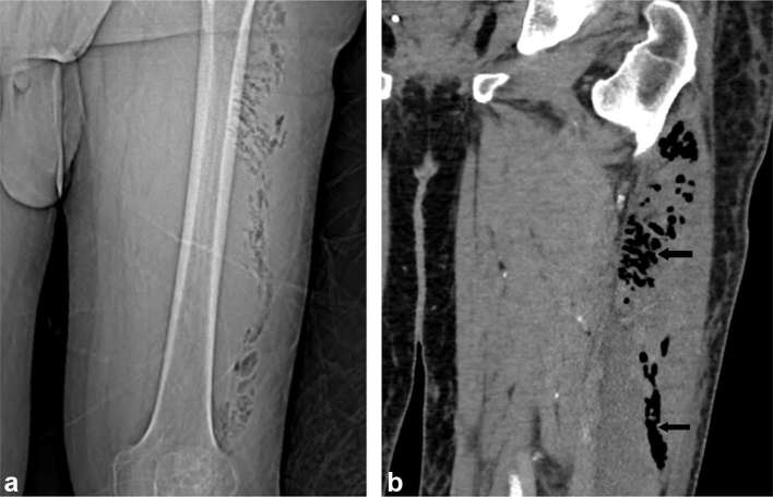
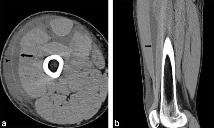
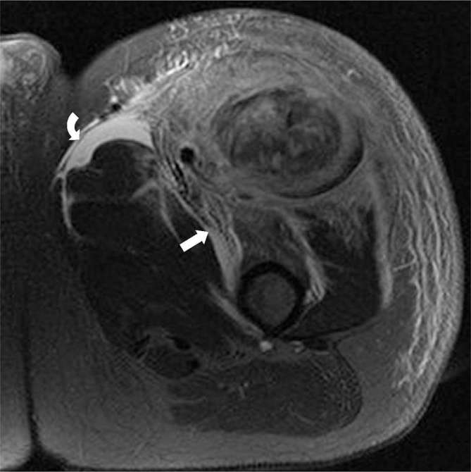
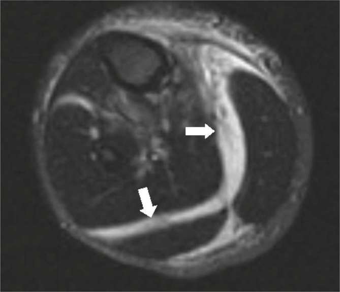
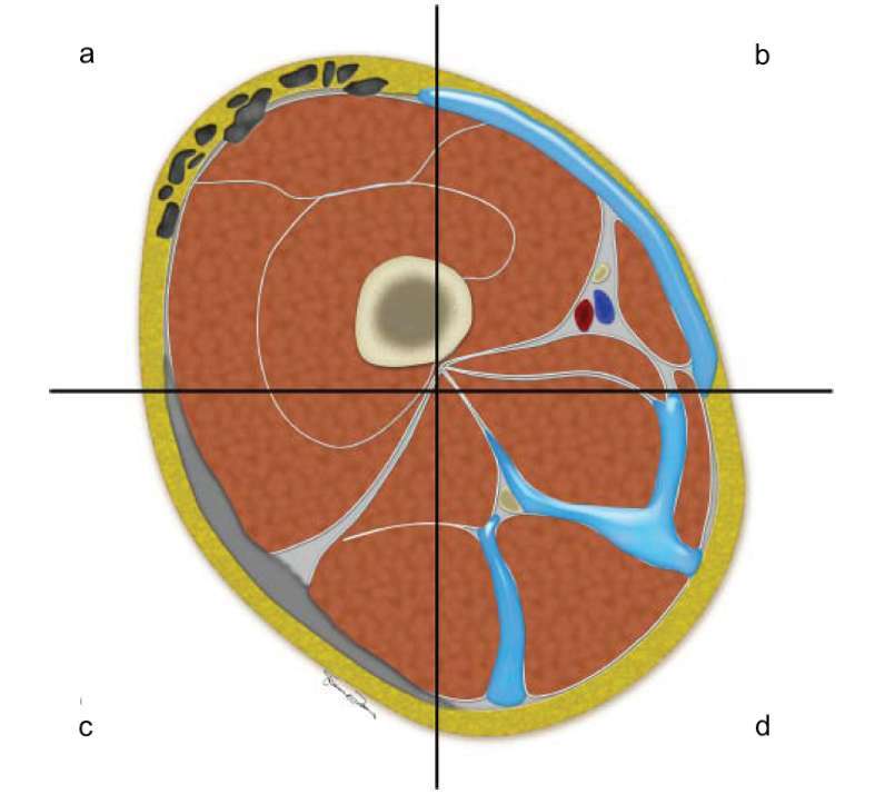
Conclusion: Although classically a clinical diagnosis, imaging is a powerful adjunct to facilitate early diagnosis in equivocal cases. Compared to plain radiography, ultrasound, CT and MR provide higher sensitivity and specificity for the diagnosis of necrotizing fasciitis. Cross-sectional imaging findings include asymmetric thickening of fascia, soft tissue air, blurring of fascial planes, inflammatory fat stranding, reactive lymphadenopathy, and nonenhancement of muscular fascia.


Figures

Figure 1.
Figure 1.
A 45-year-old male with necrotizing fasciitis of the right thigh. Extensive streaky soft-tissue gas is seen extending along the fascial planes of the right thigh on radiograph.
Figure 2.
Figure 2.
A 53-year-old male with necrotizing fasciitis of the left knee. Soft-tissue gas is seen at the lateral aspect of the left knee along the fascial planes on the radiograph.
Figure 3.
Figure 3.
A 57-year-old diabetic male with pneumoscrotum. Large volume of gas seen within the scrotum wall and scrotum sac on the scout image (curved black arrow), consistent with Fournier’s gangrene.
Figure 4.
Figure 4.
A 39-year-old male with necrotizing fasciitis of the right ankle. There is subcutaneous emphysema (arrows) overlying the right ankle with plate and screw fixation seen (a). The soft-tissue air deep to the fascia is seen as multiple echogenic foci (arrows) on ultrasound study (b).
Figure 5.
Figure 5.
A 64-year-old male with Fournier’s gangrene with perforated diverticulitis. Contrast-enhanced CT demonstrates air (arrows) and edema in the scrotum, surrounding the right testicle (a). Contrast-enhanced CT demonstrates a horse-shoe shaped perirectal air collection (arrows), extending into the subcutaneous tissues of the ischiorectal fossa and medial gluteal region (b).
Figure 6.
Figure 6.
A 55-year-old male with necrotizing Fasciitis of the left thigh. Scout film (a) and contrast-enhanced CT (b) shows intramuscular pockets of gas (arrows) in the left lateral thigh.
Figure 7.
Figure 7.
A 39-year-old-male with necrotizing fasciitis of the right thigh. Contrast-enhanced CT demonstrates crescentic subfascial fluid (arrow) with fluid also seen superficial to the fascia (arrow head) and between muscle planes (a). Sagittal CT reformation demonstrates linear fluid collection (arrow) deep to the rectus femoris muscle (b).
Figure 8.
Figure 8.
A 47-year-old male with necrotizing fasciitis of the left thigh. T2 weighted image with fat saturation of the left thigh demonstrates a large area of myonecrosis within the proximal rectus femoris muscle, with extensive muscular, fascial, and subcutaneous enhancement and edema, with crescentic fascial fluid collections, predominantly around the rectus femoris and sartorius, suggestive of necrotizing fasciitis.
Figure 9.
Figure 9.
A 35-year-old male with necrotizing fasciitis of the right calf. T2 weighted images with fat saturation demonstrates extensive high signal within the intermuscular septa surrounding the gastrocnemius and soleus muscle bellies suggestive of subfascial fluid (white arrows).
Figure 10.
Figure 10.
Cross-sectional schematic diagram through the right thigh demonstrating the various findings of necrotizing fasciitis. Subcutaneous and subfacial emphysema, which are classical finding of necrotizing fasciitis (a). Subfacial fluid along the superficial fascial layers, which can be seen in early necrotizing fasciitis (b). Diffuse thickening of the superficial fascia, which can be seen in the early phase of necrotizing fasciitis (c). Fluid tracking along deep and intermuscular fascia, which can be seen in advanced cases (d).

Similar articles

Cited by

References

    1. Chaudhry AA, Baker KS, Gould ES, Gupta R. Necrotizing fasciitis and its mimics: what radiologists need to know. AJR Am J Roentgenol 2015; 204: 128–39. doi: 10.2214/AJR.14.12676 - DOI - PubMed
    1. Fayad LM, Carrino JA, Fishman EK. Musculoskeletal infection: role of CT in the emergency department. Radiographics 2007; 27: 1723–36. doi: 10.1148/rg.276075033 - DOI - PubMed
    1. Hayeri MR, Ziai P, Shehata ML, Teytelboym OM, Huang BK. Soft-tissue infections and their imaging mimics: from cellulitis to necrotizing fasciitis. Radiographics 2016; 36: 1888–910. doi: 10.1148/rg.2016160068 - DOI - PubMed
    1. Puvanendran R, Huey JC, Pasupathy S. Necrotizing fasciitis. Can Fam Physician 2009; 55: 981–7. - PMC - PubMed
    1. Wong CH, Wang YS. The diagnosis of necrotizing fasciitis. Curr Opin Infect Dis 2005; 18: 101–6. doi: 10.1097/01.qco.0000160896.74492.ea - DOI - PubMed

Publication types