An ex vivo study of automated motion artefact correction and the impact on cone beam CT image quality and interpretability
- PMID: 29537303
- PMCID: PMC6196041
- DOI: 10.1259/dmfr.20180013
An ex vivo study of automated motion artefact correction and the impact on cone beam CT image quality and interpretability
Abstract
Objectives: To assess the impact of head motion artefacts and an automated artefact-correction system on cone beam CT (CBCT) image quality and interpretability for simulated diagnostic tasks.
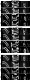
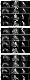
Methods: A partially dentate human skull was mounted on a robot simulating four types of head movement (anteroposterior translation, nodding, lateral rotation, and tremor), at three distances (0.75, 1.5, and 3 mm) based on two movement patterns (skull returning/not returning to the initial position). Two diagnostic tasks were simulated: dental implant planning and detection of a periapical lesion. Three CBCT units were used to examine the skull during the movements and no-motion (control): Cranex 3Dx (CRA), Orthophos SL 3D (ORT), and X1 without (X1wo) and with (X1wi) an automated motion artefact-correction system. For each diagnostic task, 88 examinations were performed. Three observers, blinded to unit and movement, scored image quality: presence of stripe artefacts (present/absent), overall unsharpness (present/absent), and image interpretability (interpretable/non-interpretable). κ statistics assessed interobserver agreement, and descriptive statistics summarized the findings.
Results: Interobserver agreement for image interpretability was good (average κ = 0.68). Regarding dental implant planning, X1wi images were interpretable by all observers, while for the other units mainly the cases with tremor were non-interpretable. Regarding detection of a periapical lesion, besides tremor, most of the 3 mm movements based on the "not returning" pattern were also non-interpretable for CRA, ORT, and X1wo. For X1wi, two observers scored 1.5 mm tremor and one observer scored 3 mm tremor as non-interpretable.
Conclusions: The automated motion artefact-correction system significantly enhanced CBCT image quality and interpretability.
Figures


Similar articles
-
Impact of movement and motion-artefact correction on image quality and interpretability in CBCT units with aligned and lateral-offset detectors.Dentomaxillofac Radiol. 2020 Jan;49(1):20190240. doi: 10.1259/dmfr.20190240. Epub 2019 Sep 24. Dentomaxillofac Radiol. 2020. PMID: 31530012 Free PMC article.
-
Impact of motion artefacts and motion-artefact correction on diagnostic accuracy of apical periodontitis in CBCT images: an ex vivo study in human cadavers.Int Endod J. 2020 Sep;53(9):1275-1288. doi: 10.1111/iej.13326. Epub 2020 Jun 11. Int Endod J. 2020. PMID: 32395820
-
Cone beam CT image artefacts related to head motion simulated by a robot skull: visual characteristics and impact on image quality.Dentomaxillofac Radiol. 2013;42(2):32310645. doi: 10.1259/dmfr/32310645. Epub 2012 Jul 27. Dentomaxillofac Radiol. 2013. PMID: 22842641 Free PMC article.
-
Image-stitching artefacts and distortion in CCD-based cephalograms and their association with sensor type and head movement: ex vivo study.Dentomaxillofac Radiol. 2020 Mar;49(3):20190315. doi: 10.1259/dmfr.20190315. Epub 2019 Nov 18. Dentomaxillofac Radiol. 2020. PMID: 31697180 Free PMC article.
-
[Metal artefact on head and neck cone-beam CT images].Fogorv Sz. 2008 Oct;101(5):171-8. Fogorv Sz. 2008. PMID: 19039918 Review. Hungarian.
Cited by
-
Objective assessment of the combined effect of exomass-related- and motion artefacts in cone beam CT.Dentomaxillofac Radiol. 2021 Jan 1;50(1):20200255. doi: 10.1259/dmfr.20200255. Epub 2020 Aug 19. Dentomaxillofac Radiol. 2021. PMID: 32706986 Free PMC article.
-
Head motion during cone-beam computed tomography: Analysis of frequency and influence on image quality.Imaging Sci Dent. 2020 Sep;50(3):227-236. doi: 10.5624/isd.2020.50.3.227. Epub 2020 Sep 16. Imaging Sci Dent. 2020. PMID: 33005580 Free PMC article.
-
Head motion and perception of discomfort by young children during simulated CBCT examinations.Dentomaxillofac Radiol. 2021 Mar 1;50(3):20200445. doi: 10.1259/dmfr.20200445. Epub 2020 Oct 30. Dentomaxillofac Radiol. 2021. PMID: 33125282 Free PMC article. Clinical Trial.
-
The Effect of Changes in the Angular Position of Implants on Metal Artifact Reduction in Cone-Beam Computed Tomography Images: A Scoping Review.Radiol Res Pract. 2023 Jul 31;2023:5539719. doi: 10.1155/2023/5539719. eCollection 2023. Radiol Res Pract. 2023. PMID: 37554657 Free PMC article.
-
Diagnostic performance of cone-beam computed tomography for scaphoid fractures: a systematic review and diagnostic meta-analysis.Sci Rep. 2021 Jan 28;11(1):2587. doi: 10.1038/s41598-021-82351-9. Sci Rep. 2021. PMID: 33510347 Free PMC article.
References
MeSH terms
LinkOut - more resources
Full Text Sources
Other Literature Sources

