

Impaired decision-making and functional neuronal network activity in systemic lupus erythematosus
- PMID: 29537670
- PMCID: PMC6282848
- DOI: 10.1002/jmri.26006
Impaired decision-making and functional neuronal network activity in systemic lupus erythematosus
Abstract
Background: Systemic lupus erythematosus (SLE) is associated with cognitive deficit but the exact neural mechanisms remain unclear.
Purpose: To explore sequential brain activities using functional magnetic resonance imaging (fMRI) during the performance of a decision-making task, and to determine whether serum or clinical markers can reflect the involvement of the brain in SLE.
Subjects: Sixteen female SLE patients without overt clinical neuropsychiatric symptoms and 16 healthy controls were included.
Field strength/sequence: 1.5T, T1 -weighted anatomic images, gradient-echo echo-planar imaging sequence, and 3D images.
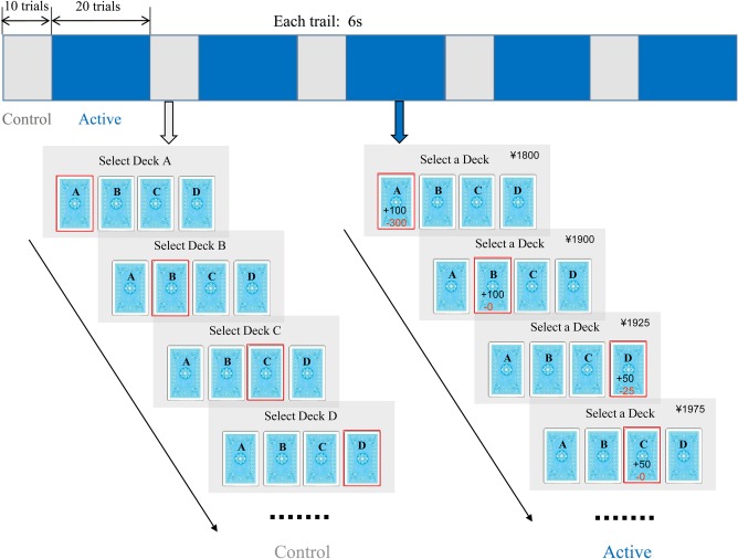
Assessment: The computer-based Iowa Gambling Task (IGT) for assessing decision-making was performed by SLE patients and 16 matched controls; brain activity was recorded via blood oxygen level-dependent (BOLD) fMRI. The amplitudes of the average BOLD responses were calculated for each individual subject, and activation data from fMRI experiments were compared between the two groups.
Statistical tests: Two-sample t-test; repeated-measures analysis of variance (ANOVA); linear regression analyses.
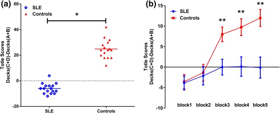
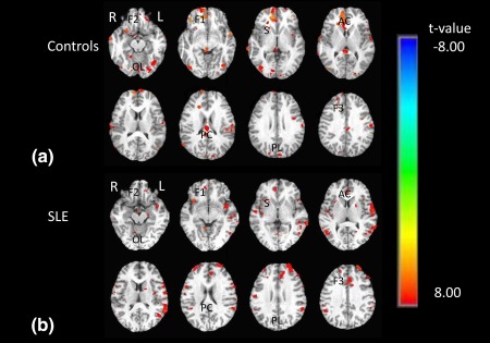
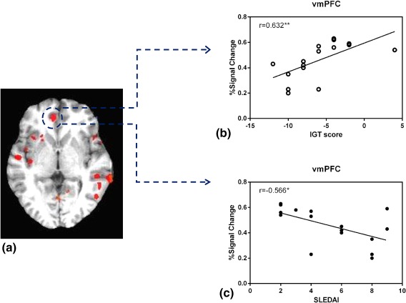
Results: Imaging revealed activity in a distributed network of brain regions in both groups, including the ventromedial prefrontal cortex (vmPFC), the orbitofrontal cortex (OFC), the dorsolateral prefrontal cortex (dlPFC), the anterior cingulate cortex (ACC), the posterior cingulate cortex (PCC), and the striatum, as well as the insular, parietal, and occipital cortices. Compared to controls, SLE patients showed lower activation in a convergence zone and the limbic system, namely, the OFC, vmPFC, ACC, and PCC, but greater activation in memory, emotion, and behavior systems involving the dlPFC, the insular cortex and the striatum. Furthermore, brain activation in the vmPFC was positively correlated with IGT scores (r = 0.63, P < 0.001), but inversely related to disease activity (r = -0.57, P < 0.01).
Data conclusion: The dynamics among the aforementioned neural systems (some hyperfunctioning, others hypofunctioning) may shed some light on the pathologic mechanisms underlying SLE without overt clinical neuropsychiatric symptoms. In addition, disease activity may potentially be used as an effective biomarker reflecting cerebral involvement in SLE.
Level of evidence: 1 Technical Efficacy: Stage 3 J. Magn. Reson. Imaging 2018;48:1508-1517.
Keywords: Iowa Gambling Task; cognitive deficit; decision-making; functional imaging; systemic lupus erythematosus.
© 2018 The Authors Journal of Magnetic Resonance Imaging published by Wiley Periodicals, Inc. on behalf of International Society for Magnetic Resonance in Medicine.
Figures
Similar articles
-
Spatial Working Memory Impairment in Patients with Non-neuropsychiatric Systemic Lupus Erythematosus: A Blood-oxygen-level Dependent Functional Magnetic Resonance Imaging Study.J Rheumatol. 2017 Feb;44(2):201-208. doi: 10.3899/jrheum.160290. Epub 2017 Jan 15. J Rheumatol. 2017. PMID: 28089970
-
Learning and memory-related brain activity dynamics are altered in systemic lupus erythematosus: a functional magnetic resonance imaging study.Lupus. 2013 May;22(6):562-73. doi: 10.1177/0961203313480399. Epub 2013 Mar 27. Lupus. 2013. PMID: 23535531
-
Decision-making in primary onset middle-age type 2 diabetes mellitus: a BOLD-fMRI study.Sci Rep. 2017 Aug 31;7(1):10246. doi: 10.1038/s41598-017-10228-x. Sci Rep. 2017. PMID: 28860463 Free PMC article.
-
[Decision-making and schizophrenia].Encephale. 2011 Dec;37 Suppl 2:S110-6. doi: 10.1016/S0013-7006(11)70036-7. Encephale. 2011. PMID: 22212839 Review. French.
-
Cognitive dysfunction and functional magnetic resonance imaging in systemic lupus erythematosus.Lupus. 2015 Oct;24(12):1239-47. doi: 10.1177/0961203315593819. Epub 2015 Jun 29. Lupus. 2015. PMID: 26124237 Review.
Cited by
-
Recent advances in the diagnosis and management of neuropsychiatric lupus.Nat Rev Rheumatol. 2024 Nov;20(11):712-728. doi: 10.1038/s41584-024-01163-z. Epub 2024 Oct 2. Nat Rev Rheumatol. 2024. PMID: 39358609 Review.
-
Altered Temporal Dynamics of Brain Activity in Multiple-Frequency Bands in Non-Neuropsychiatric Systemic Lupus Erythematosus Patients with Inactive Disease.Neuropsychiatr Dis Treat. 2021 May 7;17:1385-1395. doi: 10.2147/NDT.S292302. eCollection 2021. Neuropsychiatr Dis Treat. 2021. PMID: 33994788 Free PMC article.
-
Different patterns of cerebral perfusion in SLE patients with and without neuropsychiatric manifestations.Hum Brain Mapp. 2020 Feb 15;41(3):755-766. doi: 10.1002/hbm.24837. Epub 2019 Oct 24. Hum Brain Mapp. 2020. PMID: 31650651 Free PMC article.
-
Animal models of neuropsychiatric systemic lupus erythematosus: deciphering the complexity and guiding therapeutic development.Autoimmunity. 2024 Dec;57(1):2330387. doi: 10.1080/08916934.2024.2330387. Epub 2024 Mar 31. Autoimmunity. 2024. PMID: 38555866 Review.
-
Functional dysconnectivity and microstructural impairment of the cortico-thalamo-cortical network in women with rheumatoid arthritis: A multimodal MRI study.Heliyon. 2024 Jan 18;10(2):e24725. doi: 10.1016/j.heliyon.2024.e24725. eCollection 2024 Jan 30. Heliyon. 2024. PMID: 38304809 Free PMC article.
References
-
- Rahman A, Isenberg DA. Systemic lupus erythematosus. N Engl J Med 2008;358:929–939. - PubMed
-
- [No authors listed.] The American College of Rheumatology nomenclature and case definitions for neuropsychiatric lupus syndromes. Arthritis Rheum 1999;42:599–608. - PubMed
-
- Kozora E, Ellison MC, West S. Reliability and validity of the proposed American College of Rheumatology neuropsychological battery for systemic lupus erythematosus. Arthritis Rheum 2004;51:810–818. - PubMed
-
- Hanly JG, Fisk JD, Sherwood G, et al. Cognitive impairment in patients with systemic lupus erythematosus. J Rheumatol 1992;19:562–567. - PubMed
-
- Kozora E, Arciniegas DB, Filley CM, et al. Cognitive and neurologic status in patients with systemic lupus erythematosus without major neuropsychiatric syndromes. Arthritis Rheum 2008;59:1639–1646. - PubMed
Publication types
MeSH terms
Grants and funding
- 81774395/National Natural Science Foundation of China/International
- 81373745/National Natural Science Foundation of China/International
- 81072905/National Natural Science Foundation of China/International
- 2017A020215060/Science and Technology Planning Project of Guangdong Province of China/International
- 2009B030801323/Science and Technology Planning Project of Guangdong Province of China/International
- 2010B031600023/Science and Technology Planning Project of Guangdong Province of China/International
- S2011010005019/Natural Science Foundation of Guangdong Province of China/International
- 10151503102000015/Natural Science Foundation of Guangdong Province of China/International
- [2011] 46/Shantou Technology Bureau Science Foundation of China, Shantou Government Technology/International
- [2006] 85/Shantou Technology Bureau Science Foundation of China, Shantou Government Technology/International
LinkOut - more resources
Full Text Sources
Other Literature Sources
Medical
Research Materials
