. 2018 Mar 14;16(3):e2003586.
doi: 10.1371/journal.pbio.2003586. eCollection 2018 Mar.

Recurrent excitation between motoneurones propagates across segments and is purely glutamatergic

Affiliations

Recurrent excitation between motoneurones propagates across segments and is purely glutamatergic

Gardave S Bhumbra et al. PLoS Biol. .

Abstract

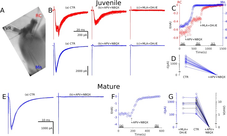
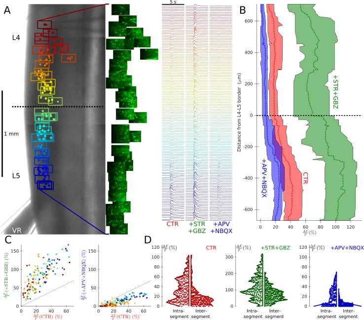
Spinal motoneurones (Mns) constitute the final output for the execution of motor tasks. In addition to innervating muscles, Mns project excitatory collateral connections to Renshaw cells (RCs) and other Mns, but the latter have received little attention. We show that Mns receive strong synaptic input from other Mns throughout development and into maturity, with fast-type Mns systematically receiving greater recurrent excitation than slow-type Mns. Optical recordings show that activation of Mns in one spinal segment can propagate to adjacent segments even in the presence of intact recurrent inhibition. While it is known that transmission at the neuromuscular junction is purely cholinergic and RCs are excited through both acetylcholine and glutamate receptors, here we show that neurotransmission between Mns is purely glutamatergic, indicating that synaptic transmission systems are differentiated at different postsynaptic targets of Mns.


Conflict of interest statement

The authors have declared that no competing interests exist.

Figures

Fig 1
Fig 1. Paired recordings from Mns showed small unitary currents that were purely glutamatergic.
Prior intramuscular injection of gastrocneumius with CTB-Alexa-Fluor-555 fluorescently labelled the dorsal motor column (A) within the coronal preparation (left) for simultaneous visualisation of Mns using infrared (middle) and confocal (right) optics. Presynaptic cells were stimulated in loose cell-attached voltage clamp (B, upper trace) while recording evoked postsynaptic responses in whole-cell voltage clamp (B, lower trace). Connected Mns were usually within 150 μM of one another, and in most paired recordings (14/18), their respective locations were recorded (group data tabulated in S1 Data). Graph C plots the relative position of each presynaptic Mn in relation to the postsynaptic cell, with colour-coded size of the corresponding reponses. A similar colour code and scale is used in graph D, showing decay time (td) against the rise time (tr). Using oblique slice preparations (see text), we investigated the pharmacology of the synapse. Panel E shows a representative recording in control (left) and following glutamatergic blockade (E, right) using APV and NBQX. The time course of changes in evoked responses during the bath application of the antagonists showed complete suppression of currents (F, top). Similar effects were observed for all 4 paired recordings (F, bottom). APV, D-2-amino-5-phosphonopentanoic acid; CTR, control conditions; Mn, motoneurone; NBQX, 1,2,3,4-tetrahydrobenzo(f)quinoxaline-7-sulphonamide.
Fig 2
Fig 2. Electrophysiological recordings from both juvenile and mature preparations showed that recurrent excitation in Mns is purely glutamatergic.
In oblique slice preparations (A) a suction electrode was used to stimulate the VR while recording respones from an Mn and, in the illustrated example, also from an RC. Note that the RC response shown includes a second component originating from a gap junction with a neighbouring RC. Panel B illustrates postsynaptic responses (stimulus artefacts truncated) of the RC (red, top) and Mn (blue, bottom) in control (left), in the presence of glutamatergic antagonists (middle) and following block of cholinergic transmission with MLA and DHβE (right). The time course of changes in currents (C, top) during application of the antagonists showed partial attenuation of RC responses and full suppression of Mn responses in the presence of glutamatergic antagonists. RC currents were only abolished by block of cholinergic receptors. In all Mns tested, the rEPSC was completely abolished by glutamatergic antagonists (D, group data tabulated in S1 Data). Similar experiments were performed on mature preparations in which Mn responses to VR stimulation were recorded in control (E, left) and during glutamatergic blockade (E, right). Once again, the time course of changes in reponses (F) showed complete suppression of Mn responses in the presence of glutamatergic antagonists. Graph G summarises the group data from Mns recorded in voltage clamp (black) and current clamp (blue) showing no effect following bath application of cholinergic antagonists, whereas glutamatergic blockade completely abolishes responses. APV, D-2-amino-5-phosphonopentanoic acid; CTR, control conditions; DHβE, dihydro-β-erythroidine; MLA, methyllycaconitine; Mn, motoneurone; RC, Renshaw cell; rEPSC, recurrent Excitatory Postsynaptic Current; VR, ventral root.
Fig 3
Fig 3. Recordings from coronal preparations showed that the magnitude of rEPSCS was related not to the position of the Mn but to its firing type.
Example traces of evoked responses (stimulus artefacts truncated) of Mns located in L5 (A, left) and L4 (A, right) segments are illustrated following stimulation of the L5 (A, top, blue) and L4 (A, bottom, red) VR; the response in the top right trace exhibited a late component, suggesting activation of a disynaptic pathway. Panel B shows the size of rEPSCs recorded from all Mns (group data tabulated in S1 Data) against their distance from L4–L5 border (rostral positive) colour-coded according to whether L5 (blue) or L4 (red) VR was stimulated using crosses for control and circles in the presence of strychnine and gabazine. There was no systematic association between rEPSCs and position as shown in the box and whisker plot (B, right) comparing intrasegmental to intersegmental responses or when comparing intersegmental responses recorded in the presence of inhibitory antagonists with control conditions. Two Mn cell types were distinguished using current clamp recordings on the basis of whether at rheobase, positive current application elicited delayed (C, left, purple), or immediate (C, right, green) firing. Delayed-firing cells (purple) were associated with a high rheobase (D, left), an accelerating initial firing rate (D, middle), and large evoked rEPSCs (D, right) in comparison to immediate-firing cells (green). The traces in panel E illustrate representative responses to VR stimulation from a delayed-firing cell (purple, left) and immediate-firing cell (green, right) recorded in voltage clamp (top) and current clamp (bottom). Graph F shows the group data, plotting the rEPSCs and rEPSPs against cell resistance and capacitance, using grey circles to denote cells that were not identified by their firing pattern at rheobase. In all 4 cases, correlations were observed, demonstrating that responses were greater in larger Mns, which tended to be of the delayed-firing type. CTR, control conditions; GBZ, gabazine; Mn, motoneurone; rEPSC, recurrent excitatory postsynaptic current; rEPSP, recurrent excitatory postsynaptic potential; STR, strychnine; VR, ventral root.
Fig 4
Fig 4. V2a interneurones do not respond to VR stimulation.
Panel A shows the location of 42 recorded V2a interneurones, identified by the expression of EGFP. The cells labelled with numbers are shown in panel B and were recorded simultaneously with an Mn. Stimulation of a VR elicited a large current in Mns but no response in the recorded V2a interneurones. EGFP, enhanced green fluorescent protein; Mn, motoneurone; VR, ventral root.
Fig 5
Fig 5. Calcium imaging from coronal preparations showed that recurrent excitation from one segment can evoke spikes in adjacent segments with intact recurrent inhibition, but these responses are abolished by glutamatergic antagonists.
An example of such a recording is illustrated in (A), which overlays the Mn positions within optical fields colour coded by position, on top of a low-magnification image (A, left) of a coronal L4–5 preparation from a ChAT-GCaMP6s mouse in which Mns express GCaMP6s. A suction pipette was used to stimulate the L5 VR while acquiring fluorescence intensities throughout each field (A, middle). Changes in Mn fluorescence were measured and plotted against time (A, right) under CTR in the presence of pharmacological blockade of inhibition (+STR+GBZ) and addition of further antagonists to block glutamatergic neurotransmission (+APV+NBQX). The results for this experiment are summarised in graph B, in which running medians and interquartile ranges of responses are plotted for the 3 conditions against the distance from the L4–5 border (rostral positive). Recurrent excitation from L5 evoked firing in Mns throughout the L4 segment, and these responses were enhanced by inhibitory antagonists and abolished by glutamatergic blockade. These effects are illustrated by the graphs in (C), which plots the signal response under control conditions with that in the presence of antagonists of inhibition (C, left) and glutamatergic blockade (C, right), preserving the colour coding of position in the rostral–caudal axis used in panel A. Violin plots summarise the group data (tabulated in S1 Data), comparing intrasegmental and intersegmental responses, showing the distribution of the magnitude of responses from all Mns under control conditions (D, left), inhibitory blockade (D, middle; on a different y-scale), and in the additional presence of glutamatergic antagonists (D, right). APV, D-2-amino-5-phosphonopentanoic acid; ChAT, choline-acetyltransferase; CTR, control conditions; GBZ, gabazine; GCaMP6, Mn, motoneurone; NBQX, 1,2,3,4-tetrahydrobenzo(f)quinoxaline-7-sulphonamide; STR, strychnine; VR, ventral root.

References

    1. Kiehn O. Decoding the organization of spinal circuits that control locomotion. Nat Rev Neurosci. 2016;17(4):224–38. doi: 10.1038/nrn.2016.9 - DOI - PMC - PubMed
    1. McCrea DA, Rybak IA. Organization of mammalian locomotor rhythm and pattern generation. Brain ResRev. 2008;57(1):134–146. - PMC - PubMed
    1. Cullheim S, Kellerth JO, Conradi S. Evidence for direct synaptic interconnections between cat spinal alpha-motoneurons via the recurrent axon collaterals: a morphological study using intracellular injection of horseradish peroxidase. Brain Res. 1977;132(1):1–10. - PubMed
    1. Gogan P, Gueritaud JP, Horcholle-Bossavit G, Tyc-Dumont S. Direct excitatory interactions between spinal motoneurones of the cat. J Physiol. 1977;272(3):755–767. - PMC - PubMed
    1. Perrins R, Roberts A. Cholinergic and electrical synapses between synergistic spinal motoneurones in the Xenopus laevis embryo. J Physiol. 1995;485 Pt 1):135–44. - PMC - PubMed

Publication types