

Cardioprotection of tilianin ameliorates myocardial ischemia-reperfusion injury: Role of the apoptotic signaling pathway
- PMID: 29538428
- PMCID: PMC5851616
- DOI: 10.1371/journal.pone.0193845
Cardioprotection of tilianin ameliorates myocardial ischemia-reperfusion injury: Role of the apoptotic signaling pathway
Abstract
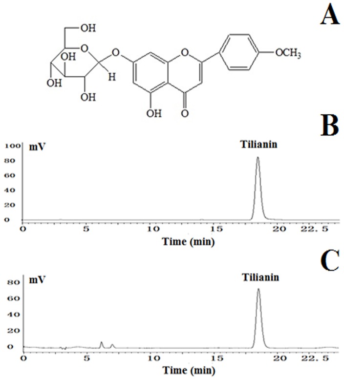
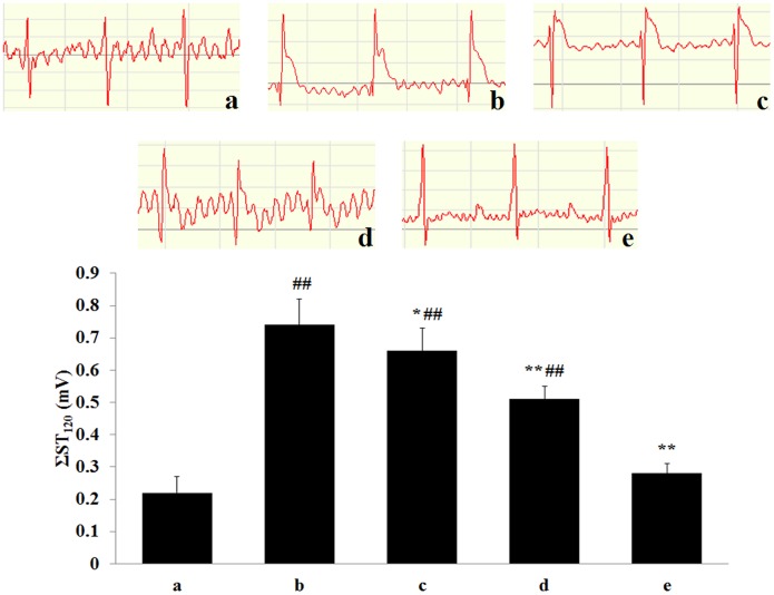
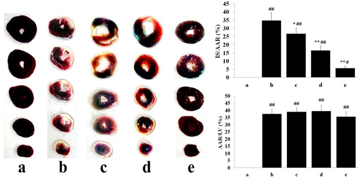
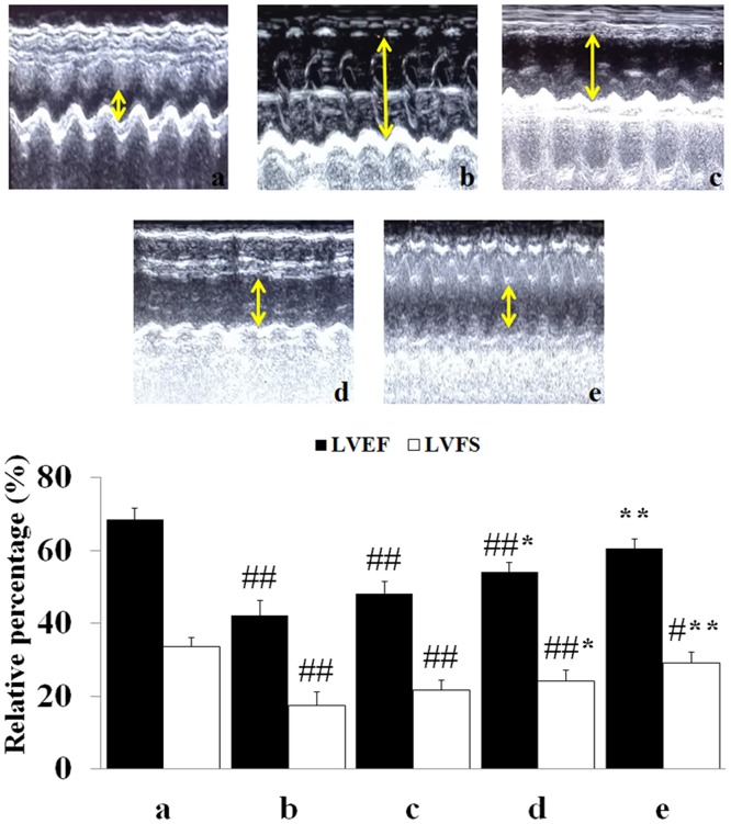
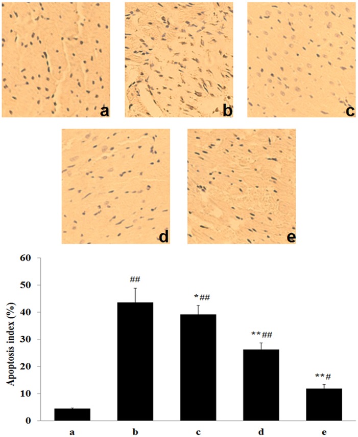
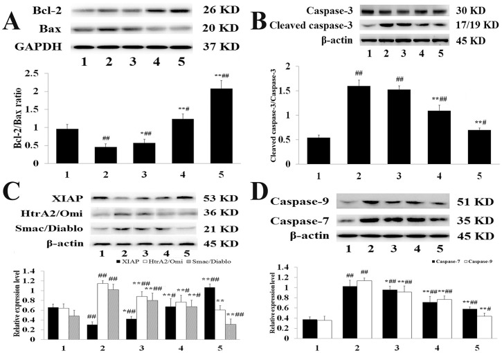
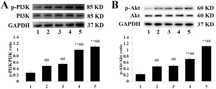
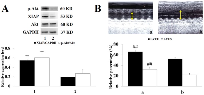
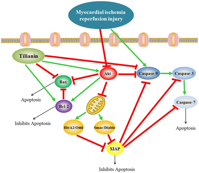
Our previous research demonstrated that tilianin protects the myocardium in a myocardial ischemia reperfusion injury (MIRI) rat model and has prominent pharmacological potential as a cardiovascular drug. Our study aimed to investigate the molecular signaling implicated in the improvement of myocardial survival induced by tilianin, a flavonoid antioxidant. Tilianin (2.5, 5, and 10 mg/kg/d) or saline was orally administered to rats for 14 days. On the 15th day, ischemia was induced by ligating the left anterior descending artery for 45 min, followed by 4 h of reperfusion. The levels of MIRI-induced serum myocardial enzymes and cardiomyocyte apoptosis as well as infarct size were examined to assess the cardioprotective effects. Cardiac tissues were collected for western blot analyses to determine the protein expression of anti-apoptotic signaling molecules. In MIRI-treated rats, our results revealed that pre-administration of high dose-tilianin the reduced release of LDH, MDA, and CK-MB and increased the plasma SOD level, and significantly attenuated the infarct size. Western blot analysis showed that a remarkable rise in expression of Bcl-2 and XIAP, and decline in expression of Bax, Smac/Diablo, HtrA2/Omi, cleaved caspase-3, caspase-7 and caspase-9 was observed in the myocardium. The apoptosis index of cardiomyocytes further supports the cardioprotective effect of tilianin. Additionally, compared with the MIRI model group, pretreatment with high dose-tilianin group upregulated phosphorylated Akt and PI3K. In contrast, using the PI3K inhibitor LY294002 to block Akt activation effectively inhibited the protective effects of tilianin against MIRI. Tilianin pretreatment was beneficial for activating the PI3K/Akt signaling pathway and inhibiting myocardial apoptosis.
Conflict of interest statement
Figures
Similar articles
-
Cardioprotective effect of breviscapine: inhibition of apoptosis in H9c2 cardiomyocytes via the PI3K/Akt/eNOS pathway following simulated ischemia/reperfusion injury.Pharmazie. 2015 Sep;70(9):593-7. Pharmazie. 2015. PMID: 26492644
-
Cardioprotective effects of tilianin in rat myocardial ischemia-reperfusion injury.Mol Med Rep. 2015 Mar;11(3):2227-33. doi: 10.3892/mmr.2014.2954. Epub 2014 Nov 14. Mol Med Rep. 2015. PMID: 25405380
-
Syringic acid mitigates myocardial ischemia reperfusion injury by activating the PI3K/Akt/GSK-3β signaling pathway.Biochem Biophys Res Commun. 2020 Oct 15;531(2):242-249. doi: 10.1016/j.bbrc.2020.07.047. Epub 2020 Aug 12. Biochem Biophys Res Commun. 2020. PMID: 32798018
-
The role of PI3K/AKT signaling pathway in myocardial ischemia-reperfusion injury.Int Immunopharmacol. 2023 Oct;123:110714. doi: 10.1016/j.intimp.2023.110714. Epub 2023 Jul 29. Int Immunopharmacol. 2023. PMID: 37523969 Review.
-
The biological and pharmacological roles of polyphenol flavonoid tilianin.Eur J Pharmacol. 2019 Jan 5;842:291-297. doi: 10.1016/j.ejphar.2018.10.044. Epub 2018 Oct 31. Eur J Pharmacol. 2019. PMID: 30389634 Review.
Cited by
-
Icariside II ameliorates myocardial ischemia and reperfusion injury by attenuating inflammation and apoptosis through the regulation of the PI3K/AKT signaling pathway.Mol Med Rep. 2020 Oct;22(4):3151-3160. doi: 10.3892/mmr.2020.11396. Epub 2020 Jul 31. Mol Med Rep. 2020. PMID: 32945440 Free PMC article.
-
Research progress on programmed cell death of cardiomyocytes in pressure-overload hypertrophic cardiomyopathy.Apoptosis. 2025 Aug 14. doi: 10.1007/s10495-025-02146-5. Online ahead of print. Apoptosis. 2025. PMID: 40813538 Review.
-
TMT-based quantitative proteomics analysis of the effects of Jiawei Danshen decoction myocardial ischemia-reperfusion injury.Proteome Sci. 2022 Dec 14;20(1):17. doi: 10.1186/s12953-022-00200-7. Proteome Sci. 2022. PMID: 36517846 Free PMC article.
-
Protective effect of luteolin on skin ischemia-reperfusion injury through an AKT-dependent mechanism.Int J Mol Med. 2018 Dec;42(6):3073-3082. doi: 10.3892/ijmm.2018.3915. Epub 2018 Oct 2. Int J Mol Med. 2018. PMID: 30280183 Free PMC article.
-
Flavonoids and Mitochondria: Activation of Cytoprotective Pathways?Molecules. 2020 Jul 4;25(13):3060. doi: 10.3390/molecules25133060. Molecules. 2020. PMID: 32635481 Free PMC article. Review.
References
-
- Kalogeris T, Baines CP, Krenz M, Korthuis RJ. Ischemia/Reperfusion. Compr Physiol. 2016. December 6; 7(1): 113–170. doi: 10.1002/cphy.c160006 - DOI - PMC - PubMed
-
- Jankovic N, Geelen A, Streppel MT, de Groot LC, Kiefte-de Jong JC, Orfanos P, et al. WHO guidelines for a healthy diet and mortality from cardiovascular disease in European and American elderly: the CHANCES project. Am J Clin Nutr. 2015. October; 102(4): 745–756. doi: 10.3945/ajcn.114.095117 - DOI - PMC - PubMed
-
- Meng G, Wang J, Xiao Y, Bai W, Xie L, Shan L, et al. GYY4137 protects against myocardial ischemia and reperfusion injury by attenuating oxidative stress and apoptosis in rats. J Biomed Res. 2015. May; 29(3): 203–213. doi: 10.7555/JBR.28.20140037 - DOI - PMC - PubMed
-
- Zhang G, Gao S, Li X, Zhang L, Tan H, Xu L, et al. Pharmacological postconditioning with lactic acid and hydrogen rich saline alleviates myocardial reperfusion injury in rats. Sci Rep. 2015. April 30; 5: 9858 doi: 10.1038/srep09858 - DOI - PMC - PubMed
-
- Law CH, Li JM, Chou HC, Chen YH, Chan HL. Hyaluronic acid-dependent protection in H9C2 cardiomyocytes: a cell model of heart ischemia-reperfusion injury and treatment. Toxocology. 2013. January 7; 303: 54–71. - PubMed
Publication types
MeSH terms
Substances
LinkOut - more resources
Full Text Sources
Other Literature Sources
Research Materials
Miscellaneous
