. 2018 Mar 14;13(3):e0194206.
doi: 10.1371/journal.pone.0194206. eCollection 2018.

Tis21-gene therapy inhibits medulloblastoma growth in a murine allograft model

Affiliations

Tis21-gene therapy inhibits medulloblastoma growth in a murine allograft model

Dario Presutti et al. PLoS One. .

Abstract

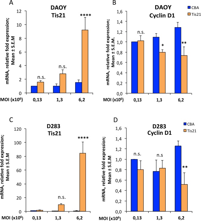
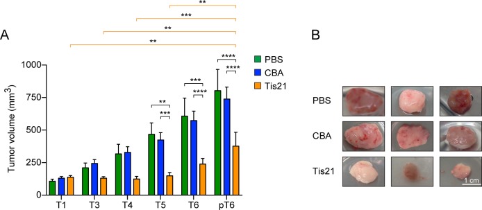
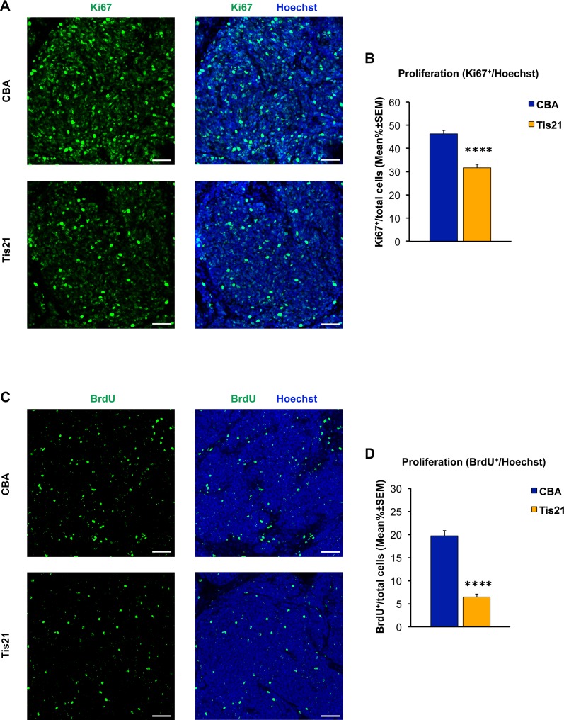
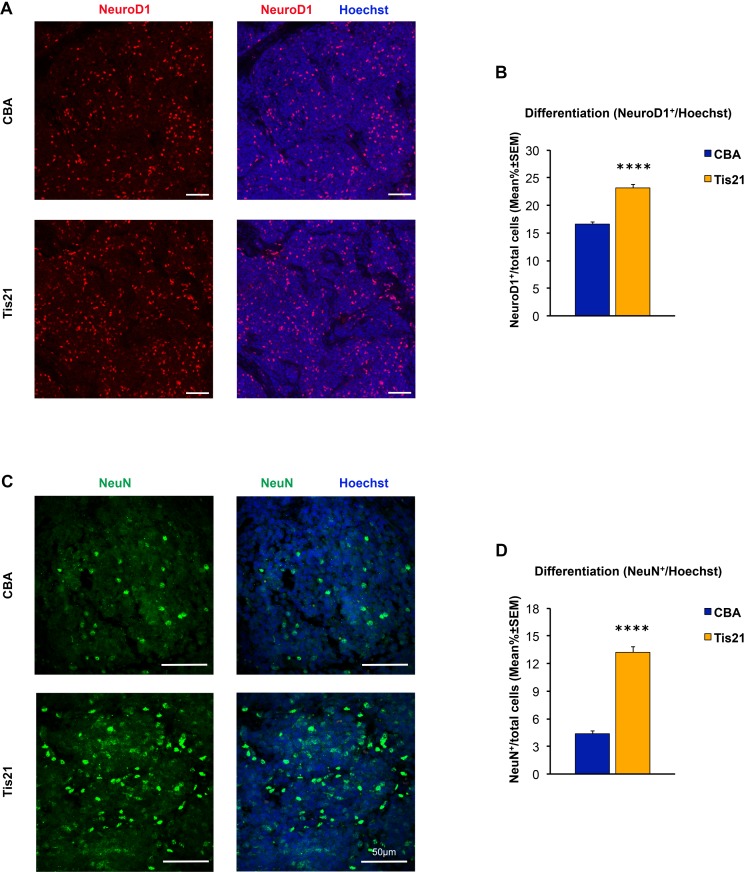
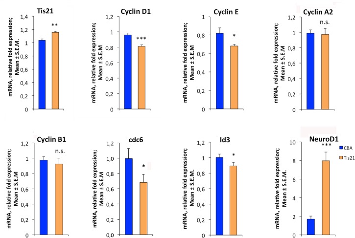
Medulloblastoma (MB), the tumor of the cerebellum, is the most frequent brain cancer in childhood and a major cause of pediatric mortality. Based on gene profiling, four MB subgroups have been identified, i.e., Wnt or Sonic Hedgehog (Shh) types, and subgroup 3 or 4. The Shh-type MB has been shown to arise from the cerebellar precursors of granule neurons (GCPs), where a hyperactivation of the Shh pathway leads to their neoplastic transformation. We have previously shown that the gene Tis21 (PC3/Btg2) inhibits the proliferation and promotes the differentiation and migration of GCPs. Moreover, the overexpression or the deletion of Tis21 in Patched1 heterozygous mice, a model of spontaneous Shh-type MB, highly reduces or increases, respectively, the frequency of MB. Here we tested whether Tis21 can inhibit MB allografts. Athymic nude mice were subcutaneously grafted with MB cells explanted from Patched1 heterozygous mice. MB allografts were then injected with adeno-associated viruses either carrying Tis21 (AAV-Tis21) or empty (AAV-CBA). We observed that the treatment with AAV-Tis21 significantly inhibited the growth of tumor nodules, as judged by their volume, and reduced the number of proliferating tumor cells (labeled with Ki67 or BrdU), relative to AAV-CBA-treated control mice. In parallel, AAV-Tis21 increased significantly tumor cells labeled with early and late neural differentiation markers. Overall the results suggest that Tis21-gene therapy slows down MB tumor growth through inhibition of proliferation and enhancement of neural differentiation. These results validate Tis21 as a relevant target for MB therapy.


Conflict of interest statement

Competing Interests: The authors have declared that no competing interests exist.

Figures

Fig 1
Fig 1. AAV-Tis21 infects DAOY and D283 MB cell lines and decreases Cyclin D1 mRNA expression levels in Tis21 dose-dependent manner.
Real-time PCR analysis of mRNA obtained from DAOY (A and B) and D283 (C and D) cells infected with AAV overexpressing Tis21 (AAV-Tis21) or AAV-CBA (control). Increasing MOI employed for infection is indicated in graphs. Average ± SEM values are from at least three independent qPCR experiments and are shown as fold change relative to the control sample (cells infected with the MOI 0.13x10⁶ of AAV-CBA), which was set to unit. Human TATA-binding protein mRNA was used as endogenous control for normalization. Two-way ANOVA followed by Fisher's PLSD test was used for the statistical analysis; * p < 0.05, ** p < 0.01, or **** p < 0.0001, n.s. p non-significant, PLSD ANOVA test.
Fig 2
Fig 2. Inhibition of Ptch1+/- MB growth in an allograft murine model by Tis21 overexpression.
A) Tumor volumes of nodules obtained by grafting primary Ptch1+/- MB tumor cells subcutaneously in nude mice flanks. The bars indicate the average ± SEM of the tumor volume at the time of the viral particles injection (T1-T6) and 24 hours after the last treatment (pT6) with PBS (green), AAV-Tis21 (orange) and AAV-CBA (blue). Groups of 3–5 mice treated with AAV-Tis21 and AAV-CBA in three independent experiments and groups of 2 mice treated with PBS in two independent experiments were analyzed. A two-way ANOVA followed by post-hoc Bonferroni's multiple comparison test was used for the statistical analysis; ** p < 0.01, *** p < 0.001, **** p < 0.0001. B) Representative images of AAV-CBA, PBS and AAV-Tis21-treated nodules are shown. Scale bars: 10 mm.
Fig 3
Fig 3. Decreased numbers of cells positive for Ki67 and BrdU in nodules of MB allograft treated with AAV-Tis21 viral particles.
A) Representative confocal images of IHC staining for the proliferation marker Ki67 (green) and for the nuclei dye Hoechst (blue). Scale bars: 50 μm. B) Quantification of the percentage ratio of cells positive for the proliferation marker Ki67 to the total number of cells (Hoechst-positive) in nodules of MB allograft treated with AAV-Tis21 (orange) and AAV-CBA (blue). **** p < 0.0001, Mann-Whitney U test; mice analyzed: n = 7 for AAV-CBA, n = 8 for AAV-Tis21; fields analyzed: n = 207 for AAV-CBA, n = 241 for AAV-Tis21. C) Representative confocal images of cells that have entered the cell cycle S-phase, either of AAV-Tis21-treated or of AAV-CBA-treated MB allograft, identified as BrdU+ cells (green). Sections are counterstained with Hoechst 33258 to visualize the nuclei. Scale bars: 50 μm. D) Quantification of proliferating cells, measured as mean ± SEM percentage ratio between number of BrdU+ cells and total number of cells (Hoechst-positive) in nodules of MB allograft treated with AAV-Tis21 (orange) and AAV-CBA (blue). **** p < 0.0001, Mann-Whitney U test; mice analyzed: n = 4 for AAV-CBA, n = 4 for AAV-Tis21; fields analyzed: n = 144 for AAV-CBA, n = 144 for AAV-Tis21.
Fig 4
Fig 4. Increased numbers of cells positive for NeuroD1 and NeuN in nodules of MB allograft treated with AAV-Tis21 viral particles.
A) Representative images of IHC staining for the differentiation marker NeuroD1 (red) and for the nuclei dye Hoechst (blue). Scale bars: 50 μm. B) Quantification of the percentage ratio of cells positive for NeuroD1 in nodules of MB allograft treated with AAV-Tis21 (orange) and AAV-CBA (blue), to the total number of cells (Hoechst-positive). **** p < 0.0001, Mann-Whitney U test. Mice analyzed: n = 7 for AAV-CBA, n = 7 for AAV-Tis21; fields analyzed: n = 201 for AAV-CBA, n = 207 for AAV-Tis21. C) Representative images of IHC staining for the differentiation marker NeuN (green). In blue the nuclei. Scale bars: 50 μm. D) Quantification of the percentage of NeuN positive cells to the total number of cells (Hoechst-positive). Orange bar (AAV-Tis21), blue bar (AAV-CBA). **** p < 0.0001, Mann-Whitney U test. Mice analyzed: n = 2 for AAV-CBA, n = 3 for AAV-Tis21; fields analyzed: n = 31 for AAV-CBA, n = 41 for AAV-Tis21.
Fig 5
Fig 5. AAV-Tis21 treatment decreases Cyclin D1 and Cyclin E and increases NeuroD1 mRNA expression levels in MB allograft tissues.
Real-time PCR analysis of RNA obtained from the nodules treated with AAV overexpressing Tis21 (AAV-Tis21) or AAV-CBA (control). Three nodules per group were analyzed. Average ± SEM values are from (at least) three independent experiments and are shown as fold change relative to the control sample (one nodule treated with AAV-CBA), which was set to unit. TATA-binding protein mRNA was used as endogenous control for normalization. * p < 0.05, ** p < 0.01, or *** p < 0.001, n.s. p non-significant, Student’s t -test.

References

    1. Smoll NR, Drummond KJ. The incidence of medulloblastomas and primitive neurectodermal tumours in adults and children. J Clin Neurosci. 2012;19: 1541–1544. doi: 10.1016/j.jocn.2012.04.009 - DOI - PubMed
    1. Brandão LA, Young Poussaint T. Posterior Fossa Tumors. Neuroimaging Clin N Am. 2017;27: 1–37. doi: 10.1016/j.nic.2016.08.001 - DOI - PubMed
    1. Rodini CO, Suzuki DE, Nakahata AM, Pereira MC, Janjoppi L, Toledo SR, et al. Aberrant signaling pathways in medulloblastomas: a stem cell connection. Arq Neuropsiquiatr. 2010;68: 947–952. - PubMed
    1. Crawford JR, MacDonald TJ, Packer RJ. Medulloblastoma in childhood: new biological advances. Lancet Neurol. 2007;6: 1073–1085. doi: 10.1016/S1474-4422(07)70289-2 - DOI - PubMed
    1. Massimino M, Biassoni V, Gandola L, Garrè ML, Gatta G, Giangaspero F, et al. Childhood medulloblastoma. Crit Rev Oncol Hematol. 2016;105: 35–51. doi: 10.1016/j.critrevonc.2016.05.012 - DOI - PubMed

Publication types

MeSH terms

Substances