

Role of Toll-like receptors and interferon regulatory factors in different experimental heart failure models of diverse etiology: IRF7 as novel cardiovascular stress-inducible factor
- PMID: 29538462
- PMCID: PMC5851607
- DOI: 10.1371/journal.pone.0193844
Role of Toll-like receptors and interferon regulatory factors in different experimental heart failure models of diverse etiology: IRF7 as novel cardiovascular stress-inducible factor
Abstract
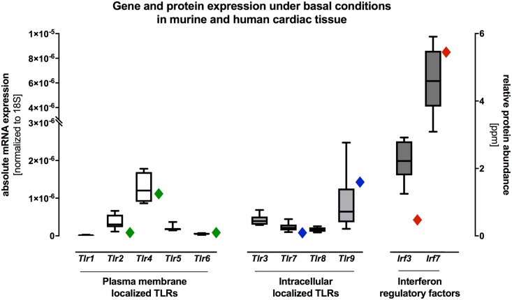
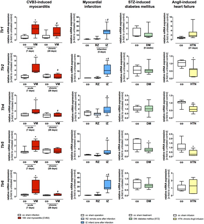
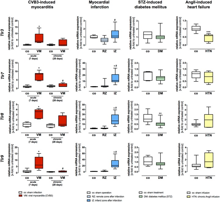
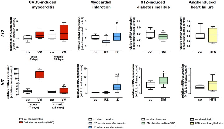
Heart failure (HF) is a leading cause of morbidity and mortality in the western world. Although optimal medical care and treatment is widely available, the prognosis of patients with HF is still poor. Toll-like receptors (TLRs) are important compartments of the innate immunity. Current studies have identified TLRs as critical mediators in cardiovascular diseases. In the present study, we investigated the involvement of TLRs and interferon (IFN) regulatory factors (IRFs) in different experimental HF models including viral myocarditis, myocardial ischemia, diabetes mellitus, and cardiac hypertrophy. In addition, we investigated for the first time comprehensive TLR and IRF gene and protein expression under basal conditions in murine and human cardiac tissue. We found that Tlr4, Tlr9 and Irf7 displayed highest gene expression under basal conditions, indicating their significant role in first-line defense in the murine and human heart. Moreover, induction of TLRs and IRFs clearly differs between the various experimental HF models of diverse etiology and the concomitant inflammatory status. In the HF model of acute viral-induced myocarditis, TLR and IRF activation displayed the uppermost gene expression in comparison to the remaining experimental HF models, indicating the highest amount of myocardial inflammation in myocarditis. In detail, Irf7 displayed by far the highest gene expression during acute viral infection. Interestingly, post myocardial infarction TLR and IRF gene expression was almost exclusively increased in the infarct zone after myocardial ischemia (Tlr2, Tlr3, Tlr6, Tlr7, Tlr9, Irf3, Irf7). With one exception, Irf3 showed a decreased gene expression in the remote zone post infarction. Finally, we identified Irf7 as novel cardiovascular stress-inducible factor in the pathologically stressed heart. These findings on TLR and IRF function in the inflamed heart highlight the complexity of inflammatory immune response and raise more interesting questions for future investigation.
Conflict of interest statement
Figures
Similar articles
-
A protective role of IRF3 and IRF7 signalling downstream TLRs in the development of vein graft disease via type I interferons.J Intern Med. 2017 Dec;282(6):522-536. doi: 10.1111/joim.12679. Epub 2017 Sep 27. J Intern Med. 2017. PMID: 28857295
-
Transcriptional regulation of lipopolysaccharide (LPS)-induced Toll-like receptor (TLR) expression in murine macrophages: role of interferon regulatory factors 1 (IRF-1) and 2 (IRF-2).J Endotoxin Res. 2006;12(5):285-95. doi: 10.1179/096805106X118834. J Endotoxin Res. 2006. PMID: 17059692 Free PMC article.
-
Molecular immune mechanisms of HPV-infected HaCaT cells in vitro based on toll-like receptors signaling pathway.J Clin Lab Anal. 2020 Mar;34(3):e23101. doi: 10.1002/jcla.23101. Epub 2019 Nov 29. J Clin Lab Anal. 2020. PMID: 31785031 Free PMC article.
-
Direct Inhibition of IRF-Dependent Transcriptional Regulatory Mechanisms Associated With Disease.Front Immunol. 2019 May 24;10:1176. doi: 10.3389/fimmu.2019.01176. eCollection 2019. Front Immunol. 2019. PMID: 31178872 Free PMC article. Review.
-
The Role of Toll-Like Receptor Signaling in the Progression of Heart Failure.Mediators Inflamm. 2018 Feb 8;2018:9874109. doi: 10.1155/2018/9874109. eCollection 2018. Mediators Inflamm. 2018. PMID: 29576748 Free PMC article. Review.
Cited by
-
Human cardiac fibroblasts produce pro-inflammatory cytokines upon TLRs and RLRs stimulation.Mol Cell Biochem. 2021 Sep;476(9):3241-3252. doi: 10.1007/s11010-021-04157-7. Epub 2021 Apr 21. Mol Cell Biochem. 2021. PMID: 33881711 Free PMC article.
-
Deep insight into cytokine storm: from pathogenesis to treatment.Signal Transduct Target Ther. 2025 Apr 16;10(1):112. doi: 10.1038/s41392-025-02178-y. Signal Transduct Target Ther. 2025. PMID: 40234407 Free PMC article. Review.
-
Sacubitril/Valsartan attenuates progression of diabetic cardiomyopathy through immunomodulation properties: an opportunity to prevent progressive disease.Cardiovasc Diabetol. 2025 May 14;24(1):206. doi: 10.1186/s12933-025-02741-5. Cardiovasc Diabetol. 2025. PMID: 40369551 Free PMC article.
-
Sex-dependent effects of genetic upregulation of activated protein C on delayed effects of acute radiation exposure in the mouse heart, small intestine, and skin.PLoS One. 2021 May 24;16(5):e0252142. doi: 10.1371/journal.pone.0252142. eCollection 2021. PLoS One. 2021. PMID: 34029348 Free PMC article.
-
Interferons and interferon-related pathways in heart disease.Front Cardiovasc Med. 2024 Apr 11;11:1357343. doi: 10.3389/fcvm.2024.1357343. eCollection 2024. Front Cardiovasc Med. 2024. PMID: 38665231 Free PMC article. Review.
References
-
- Bui AL, Horwich TB, Fonarow GC. Epidemiology and risk profile of heart failure. Nature reviews Cardiology. 2011;8(1):30–41. doi: 10.1038/nrcardio.2010.165 . - DOI - PMC - PubMed
-
- McMurray JJ, Adamopoulos S, Anker SD, Auricchio A, Bohm M, Dickstein K, et al. ESC guidelines for the diagnosis and treatment of acute and chronic heart failure 2012: The Task Force for the Diagnosis and Treatment of Acute and Chronic Heart Failure 2012 of the European Society of Cardiology. Developed in collaboration with the Heart Failure Association (HFA) of the ESC. European journal of heart failure. 2012;14(8):803–69. doi: 10.1093/eurjhf/hfs105 . - DOI - PubMed
-
- Yancy CW, Jessup M, Bozkurt B, Butler J, Casey DE Jr., Drazner MH, et al. 2013 ACCF/AHA guideline for the management of heart failure: executive summary: a report of the American College of Cardiology Foundation/American Heart Association Task Force on practice guidelines. Circulation. 2013;128(16):1810–52. doi: 10.1161/CIR.0b013e31829e8807 . - DOI - PubMed
-
- Yeung DF, Boom NK, Guo H, Lee DS, Schultz SE, Tu JV. Trends in the incidence and outcomes of heart failure in Ontario, Canada: 1997 to 2007. CMAJ: Canadian Medical Association journal = journal de l’Association medicale canadienne. 2012;184(14):E765–73. doi: 10.1503/cmaj.111958 . - DOI - PMC - PubMed
-
- Zarrinkoub R, Wettermark B, Wandell P, Mejhert M, Szulkin R, Ljunggren G, et al. The epidemiology of heart failure, based on data for 2.1 million inhabitants in Sweden. European journal of heart failure. 2013;15(9):995–1002. doi: 10.1093/eurjhf/hft064 . - DOI - PubMed
Publication types
MeSH terms
Substances
LinkOut - more resources
Full Text Sources
Other Literature Sources
Medical
Research Materials
Miscellaneous
