. 2018 Jan;110(1):44-51.
doi: 10.5935/abc.20180008.

Melatonin-Induced Protective Effects on Cardiomyocytes Against Reperfusion Injury Partly Through Modulation of IP3R and SERCA2a Via Activation of ERK1

[Article in English, Portuguese]
Affiliations

Melatonin-Induced Protective Effects on Cardiomyocytes Against Reperfusion Injury Partly Through Modulation of IP3R and SERCA2a Via Activation of ERK1

[Article in English, Portuguese]
Shunying Hu et al. Arq Bras Cardiol. 2018 Jan.

Abstract

Background: Melatonin is a neuroendocrine hormone synthesized primarily by the pineal gland that is indicated to effectively prevent myocardial reperfusion injury. It is unclear whether melatonin protects cardiac function from reperfusion injury by modulating intracellular calcium homeostasis.

Objective: Demonstrate that melatonin protect against myocardial reperfusion injury through modulating IP3R and SERCA2a to maintain calcium homeostasis via activation of ERK1 in cardiomyocytes.

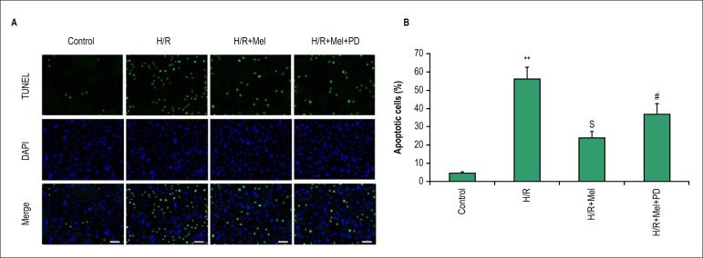
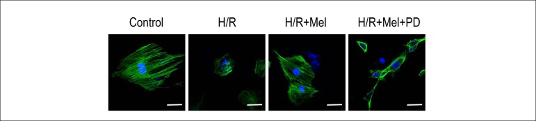
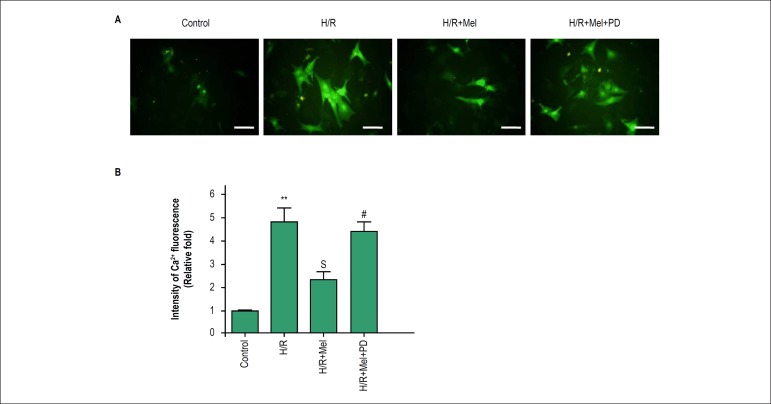
Methods: In vitro experiments were performed using H9C2 cells undergoing simulative hypoxia/reoxygenation (H/R) induction. Expression level of ERK1, IP3R and SERCA2a were assessed by Western Blots. Cardiomyocytes apoptosis was detected by TUNEL. Phalloidin-staining was used to assess alteration of actin filament organization of cardiomyocytes. Fura-2 /AM was used to measure intracellular Ca2+ concentration. Performing in vivo experiments, myocardial expression of IP3R and SERCA2a were detected by immunofluorescence staining using myocardial ischemia/ reperfusion (I/R) model in rats.

Results: In vitro results showed that melatonin induces ERK1 activation in cardiomyocytes against H/R which was inhibited by PD98059 (ERK1 inhibitor). The results showed melatonin inhibit apoptosis of cardiomyocytes and improve actin filament organization in cardiomyocytes against H/R, because both could be reversed by PD98059. Melatonin was showed to reduce calcium overload, further to inhibit IP3R expression and promote SERCA2a expression via ERK1 pathway in cardiomyocytes against H/R. Melatonin induced lower IP3R and higher SERCA2a expression in myocardium that were reversed by PD98059.

Conclusion: melatonin-induced cardioprotection against reperfusion injury is at least partly through modulation of IP3R and SERCA2a to maintain intracellular calcium homeostasis via activation of ERK1.


Conflict of interest statement

Potential Conflict of Interest

No potential conflict of interest relevant to this article was reported.

Figures

Figure 1
Figure 1
Melatonin promoted activation of ERK1 in H9C2 cells against H/R. H9C2 cells incubated in normal condition or in simulated H/R condition, in simulated H/R condition plus pretreatment with melatonin, or in simulated H/R condition plus pretreatment with melatonin and PD98059 (ERK1 inhibitor).The expression levels of phosphorylated ERK1 (p-ERK1) were evaluated by Western blotting (A) and were quantitatively analyzed (B). All values are presented as the mean±SD. n = 3. SSp < 0.01 vs. H/R group; #p < 0.05 vs. H/R+Mel group.(Control: control group; H/R:H/R group; H/R+mel: H/R+ melatonin group; H/R+mel+PD: H/R+ melatonin+PD98059 group)
Figure 2
Figure 2
Melatonin prevents H9C2 cells apoptosis against H/R via ERK1 in vitro. Apoptosis was assessed by fluorescence TUNEL. Representative TUNEL staining images (A) and quantitative analysis in H9C2 cells(B). bar = 50 µm. All values are presented as the mean±SD. n = 3.**p < 0.01 vs. control group; Sp < 0.05 vs. H/R group; #p < 0.05 vs. H/R+Mel group. (Control: control group; H/R:H/R group; H/R+mel: H/R+ melatonin group; H/R+mel+PD: H/R+ melatonin+PD98059 group)
Figure 3
Figure 3
Melatonin protects F-actin organization in H9C2 cells against H/R via ERK1 in vitro. Representative confocal microscopy images show H9C2 cells stained with FITC-phalloidin. The results showed that simulated H/R induced more diffuse and irregular actin disposition compared with control group. Melatonin preserved more regular and well-defined actin organization and PD98059 (ERK1 inhibitor) reduced the protection of melatonin. bar = 20µm. (Control: control group; H/R:H/R group; H/R+mel: H/R+ melatonin group; H/R+mel+PD: H/R+ melatonin+PD98059 group)
Figure 4
Figure 4
Melatonin reduces Ca2+ overload in H9C2 cells against H/R via ERK1 in vitro. Ca2+ content was assessed using Fura-2/AM in H9C2 cells incubated in normal condition or in simulated H/R condition, in simulated H/R condition plus pretreatment with melatonin, or in simulated H/R condition plus pretreatment with melatonin and PD98059 (ERK1 inhibitor). The green fluorescence intensity by Fura-2 was obviously stronger in H/R group, and melatonin pretreatment reversed the change which was inhibited by ERK1 inhibitor.bar = 30 µm. All values are presented as the mean ± SD. n = 3.**p < 0.01 vs. control group; Sp < 0.05 vs. H/R group; #p < 0.05 vs. H/R+Mel group. (Control: control group; H/R:H/R group; H/R+mel: H/R+ melatonin group; H/R+mel+PD: H/R+ melatonin+PD98059 group)
Figure 5
Figure 5
Melatonin modulated expression of SERCA2a and IP3R in H9C2 cells against H/R via ERK1 pathway in vitro. The results indicated melatonin inhibited expression of IP3R and promoted expression of SERCA2a which was reduced by PD98059. Representative Western blot images (A) and quantitative analysis (B-C) showed melatonin’s effect on expression of IP3R and SERCA2a via ERK1 pathway in H9C2 cells against H/R.bar = 30 µm. **p < 0.01 vs. control group; Sp < 0.05 vs. H/R group; #p < 0.05 vs. H/R+Mel group. (Control: control group; H/R:H/R group; H/R+mel: H/R+ melatonin group; H/R+mel+PD: H/R+ melatonin+PD98059 group)
Figure.6
Figure.6
Melatonin modulated expression of IP3R and SERCA2a via ERK1 pathway in reperfused rat hearts. In reperfused myocardium, expression of IP3R and SERCA2a were assessed by immunofluorescence staining (A) and quantitative analysis (B-C). The results showed that fluorescence intensity of IP3R was increased in I/R group more than in control group, but was lower in melatonin group compared with I/R group. On the contrary, melatonin increased expression of SERCA2a in reperfused myocardium. Both of the effects of melatonin on expression of IP3R and SERCA2a were inhibited by PD98059 (ERK1 inhibitor). bar = 30 µm. All values are presented as the mean ± SD. n = 5.*p < 0.05 vs. control group; Sp < 0.05 vs. I/R group; #p < 0.05 vs. I/R+Mel group. (Control: control group; I/R:I/R group; I/R+mel: I/R+ melatonin group; I/R+mel+PD: I/R+ melatonin+PD98059 group).

Comment in

References

    1. Hausenloy DJ, Yellon DM. Myocardial ischemia-reperfusion injury: a neglected therapeutic target. J Clin Invest. 2013;123(1):92–100. doi: 10.1172/JCI62874. - DOI - PMC - PubMed
    1. Morel O, Perret T, Delarche N, Labeque JN, Jouve B, Elbaz M, et al. Pharmacological approaches to reperfusion therapy. Cardiovasc Res. 2012;94(2):246–252. doi: 10.1093/cvr/cvs114. - DOI - PubMed
    1. Ibanez B, Heusch G, Ovize M, Van de Werf F. Evolving therapies for myocardial ischemia/reperfusion injury. J Am Coll Cardiol. 2015;65(14):1454–1471. doi: 10.1016/j.jacc.2015.02.032. - DOI - PubMed
    1. Chen M, Zheng YY, Song YT, Xue JY, Liang ZY, Yan XX, et al. Pretreatment with low-dose gadolinium chloride attenuates myocardial ischemia/reperfusion injury in rats. Acta Pharmacol Sin. 2016;37(4):453–462. doi: 10.1038/aps.2015.156. - DOI - PMC - PubMed
    1. Bulluck H, Yellon DM, Hausenloy DJ. Reducing myocardial infarct size: challenges and future opportunities. Heart. 2016;102(5):341–348. doi: 10.1136/heartjnl-2015-307855. - DOI - PMC - PubMed

MeSH terms

Substances