

Thrombospondin 1 Triggers Osteosarcoma Cell Metastasis and Tumor Angiogenesis
- PMID: 29540257
- PMCID: PMC7848452
- DOI: 10.3727/096504018X15208993118389
Thrombospondin 1 Triggers Osteosarcoma Cell Metastasis and Tumor Angiogenesis
Abstract
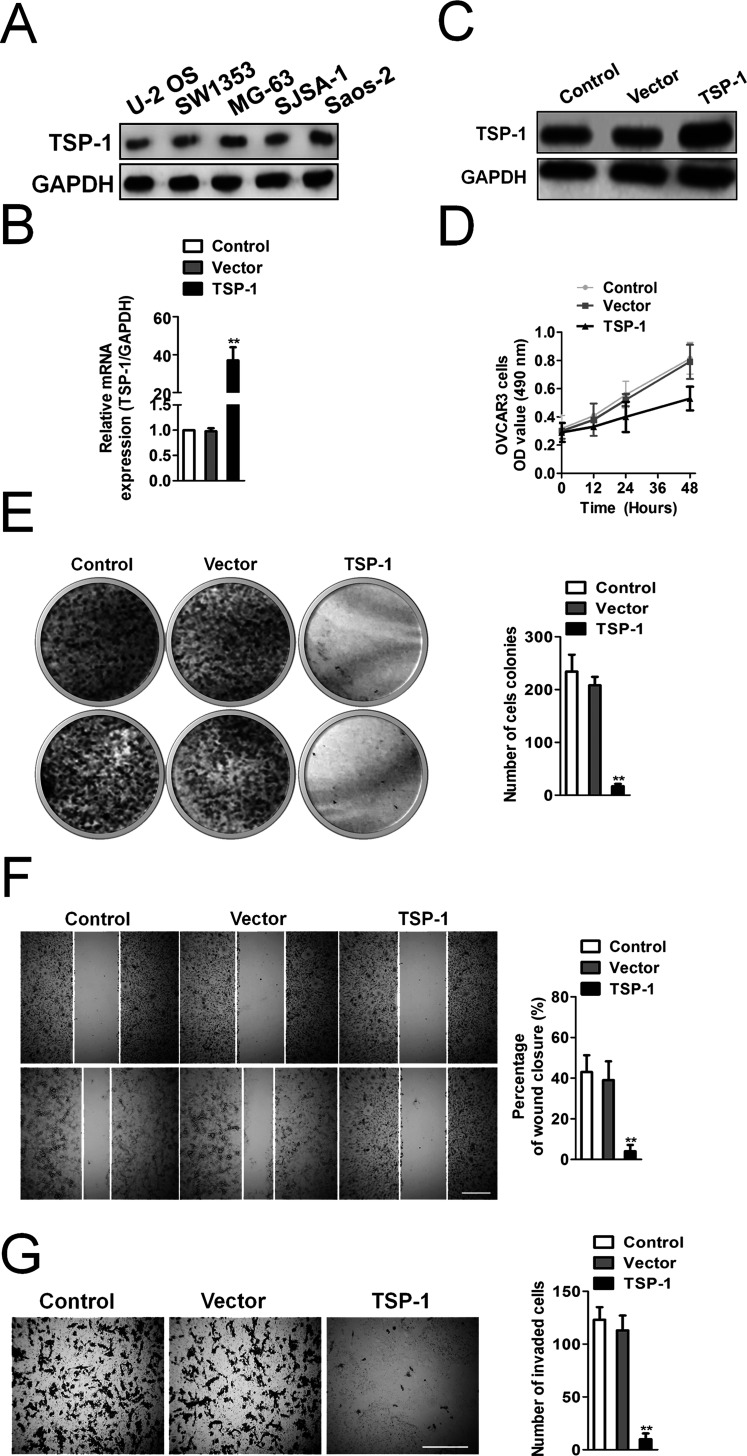
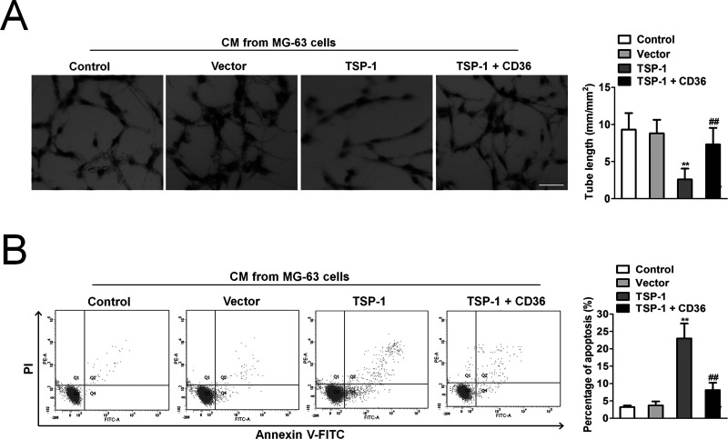
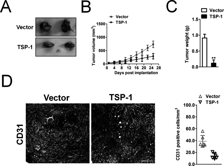
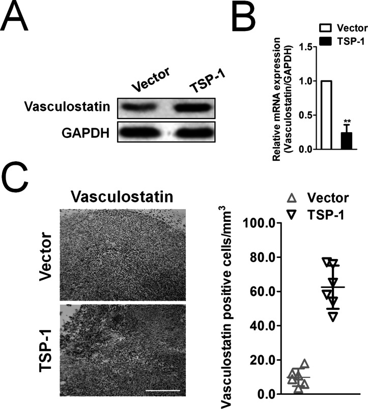
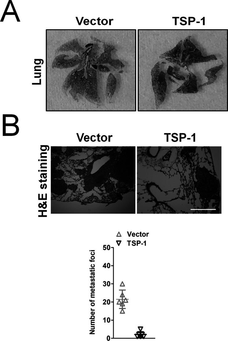
Osteosarcomas, especially those with metastatic or unresectable disease, have limited treatment options. The antitumor effects of pharmacologic inhibitors of angiogenesis in osteosarcomas are hampered in patients by the rapid development of tumor resistance, notably through increased invasiveness and accelerated metastasis. Here we demonstrated that thrombospondin 1 (TSP-1) is a potent inhibitor of the growth and metastasis of the osteosarcoma cell line MG-63. Moreover, we demonstrate that upregulation of TSP-1 facilitated expression of vasculostatin in MG-63 cells. In angiogenesis assays, overexpression of TSP-1 inhibited MG-63 cells and induced tube formation of human umbilical vein endothelial cells (HUVECs) in a CD36-dependent fashion. Finally, in xenografted tumors, we observed that TSP-1 overexpression inhibited angiogenesis and tumor growth. These results provided strong evidence for an important role of the TSP-1/CD36/vasculostatin signaling axis in mediating the antiangiogenic activity of osteosarcoma.
Conflict of interest statement
The authors declare no conflicts of interest.
Figures
Similar articles
-
CD36-mediated activation of endothelial cell apoptosis by an N-terminal recombinant fragment of thrombospondin-2 inhibits breast cancer growth and metastasis in vivo.Breast Cancer Res Treat. 2011 Jul;128(2):337-46. doi: 10.1007/s10549-010-1085-7. Epub 2010 Aug 17. Breast Cancer Res Treat. 2011. PMID: 20714802 Free PMC article.
-
Inhibition of tumor growth by systemic treatment with thrombospondin-1 peptide mimetics.Int J Cancer. 2002 Apr 10;98(5):682-9. doi: 10.1002/ijc.10247. Int J Cancer. 2002. PMID: 11920636
-
Thrombospondin-1/CD36 pathway contributes to bone marrow-derived angiogenic cell dysfunction in type 1 diabetes via Sonic hedgehog pathway suppression.Am J Physiol Endocrinol Metab. 2013 Dec;305(12):E1464-72. doi: 10.1152/ajpendo.00516.2013. Epub 2013 Oct 22. Am J Physiol Endocrinol Metab. 2013. PMID: 24148348 Free PMC article.
-
Regulation of tumor angiogenesis by thrombospondin-1.Biochim Biophys Acta. 2006 Apr;1765(2):178-88. doi: 10.1016/j.bbcan.2005.11.002. Epub 2005 Dec 21. Biochim Biophys Acta. 2006. PMID: 16406676 Review.
-
CD36: a critical anti-angiogenic receptor.Front Biosci. 2003 Sep 1;8:s874-82. doi: 10.2741/1168. Front Biosci. 2003. PMID: 12957861 Review.
Cited by
-
Ubiquitination in lipid metabolism reprogramming: implications for pediatric solid tumors.Front Immunol. 2025 Apr 30;16:1554311. doi: 10.3389/fimmu.2025.1554311. eCollection 2025. Front Immunol. 2025. PMID: 40370434 Free PMC article. Review.
-
Integrin signaling in tumor biology: mechanisms of intercellular crosstalk and emerging targeted therapies.PeerJ. 2025 May 7;13:e19328. doi: 10.7717/peerj.19328. eCollection 2025. PeerJ. 2025. PMID: 40352270 Free PMC article. Review.
-
Exosomal mRNA Cargo are biomarkers of tumor and immune cell populations in pediatric osteosarcoma.Transl Oncol. 2024 Aug;46:102008. doi: 10.1016/j.tranon.2024.102008. Epub 2024 Jun 8. Transl Oncol. 2024. PMID: 38852279 Free PMC article.
-
Deciphering the role of CD47 in cancer immunotherapy.J Adv Res. 2024 Sep;63:129-158. doi: 10.1016/j.jare.2023.10.009. Epub 2023 Oct 28. J Adv Res. 2024. PMID: 39167629 Free PMC article. Review.
-
Sphingolipid metabolism is associated with osteosarcoma metastasis and prognosis: Evidence from interaction analysis.Front Endocrinol (Lausanne). 2022 Aug 29;13:983606. doi: 10.3389/fendo.2022.983606. eCollection 2022. Front Endocrinol (Lausanne). 2022. PMID: 36105405 Free PMC article.
References
-
- Rogozhin DV, Bulycheva IV, Konovalov DM, Talalaev AG, Roshchin VY, Ektova AP, Bogoroditsky YS, Strykov VA, Kazakova AN, Olshanskaya YV, Kachanov DY, Tereshchenko GV. [Classical osteosarcoma in children and adolescent]. Arkh Patol. 2015;77(5):68–74. - PubMed
-
- Zhao GS, Gao ZR, Zhang Q, Tang XF, Lv YF, Zhang ZS, Zhang Y, Tan QL, Peng DB, Jiang DM, Guo QN. TSSC3 promotes autophagy via inactivating the Src-mediated PI3K/Akt/mTOR pathway to suppress tumorigenesis and metastasis in osteosarcoma, and predicts a favorable prognosis. J Exp Clin Cancer Res. 2018;37(1):188. - PMC - PubMed
-
- Ohba T, Cates JM, Cole HA, Slosky DA, Haro H, Ando T, Schwartz HS, Schoenecker JG. Autocrine VEGF/VEGFR1 signaling in a subpopulation of cells associates with aggressive osteosarcoma. Mol Cancer Res. 2014;12(8):1100–11. - PubMed
MeSH terms
Substances
LinkOut - more resources
Full Text Sources
Other Literature Sources
Medical
Miscellaneous