Review
. 2018 Mar 14:360:j5492.
doi: 10.1136/bmj.j5492.

Pulmonary arterial hypertension: pathogenesis and clinical management

Affiliations
Review

Pulmonary arterial hypertension: pathogenesis and clinical management

Thenappan Thenappan et al. BMJ. .

Abstract

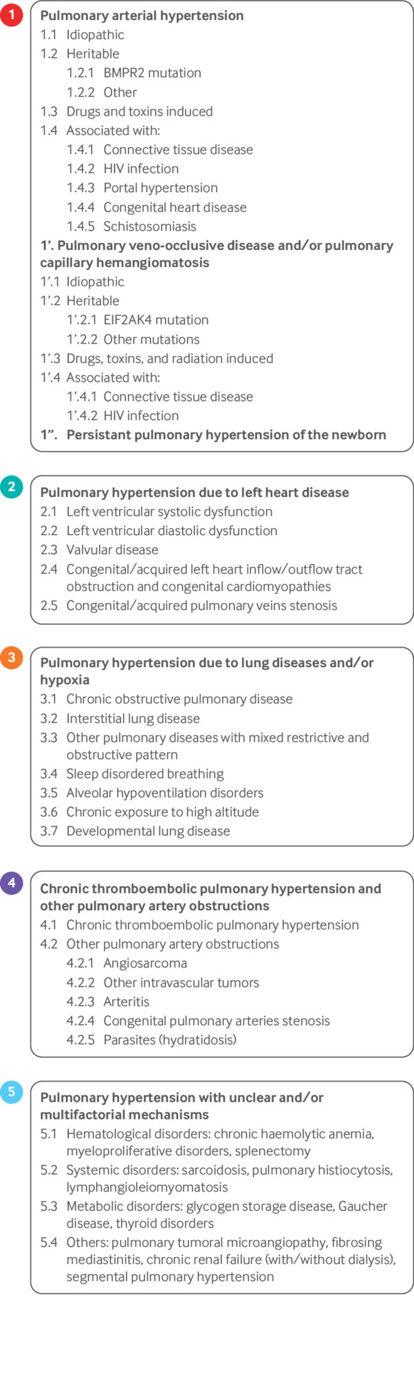
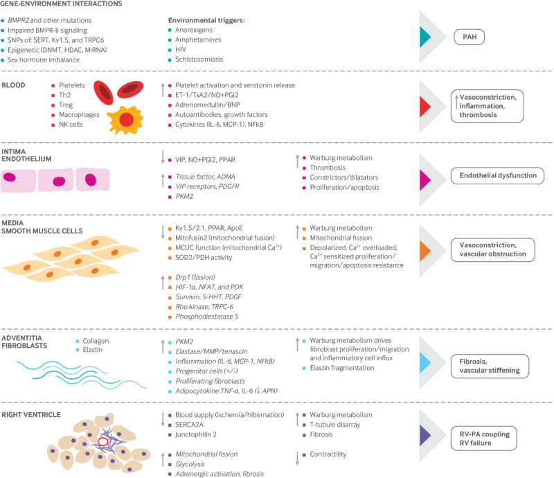
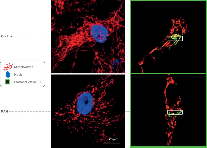
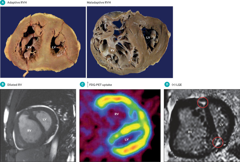
Pulmonary hypertension is defined as a resting mean pulmonary artery pressure of 25 mm Hg or above. This review deals with pulmonary arterial hypertension (PAH), a type of pulmonary hypertension that primarily affects the pulmonary vasculature. In PAH, the pulmonary vasculature is dynamically obstructed by vasoconstriction, structurally obstructed by adverse vascular remodeling, and pathologically non-compliant as a result of vascular fibrosis and stiffening. Many cell types are abnormal in PAH, including vascular cells (endothelial cells, smooth muscle cells, and fibroblasts) and inflammatory cells. Progress has been made in identifying the causes of PAH and approving new drug therapies. A cancer-like increase in cell proliferation and resistance to apoptosis reflects acquired abnormalities of mitochondrial metabolism and dynamics. Mutations in the type II bone morphogenetic protein receptor (BMPR2) gene dramatically increase the risk of developing heritable PAH. Epigenetic dysregulation of DNA methylation, histone acetylation, and microRNAs also contributes to disease pathogenesis. Aberrant bone morphogenetic protein signaling and epigenetic dysregulation in PAH promote cell proliferation in part through induction of a Warburg mitochondrial-metabolic state of uncoupled glycolysis. Complex changes in cytokines (interleukins and tumor necrosis factor), cellular immunity (T lymphocytes, natural killer cells, macrophages), and autoantibodies suggest that PAH is, in part, an autoimmune, inflammatory disease. Obstructive pulmonary vascular remodeling in PAH increases right ventricular afterload causing right ventricular hypertrophy. In some patients, maladaptive changes in the right ventricle, including ischemia and fibrosis, reduce right ventricular function and cause right ventricular failure. Patients with PAH have dyspnea, reduced exercise capacity, exertional syncope, and premature death from right ventricular failure. PAH targeted therapies (prostaglandins, phosphodiesterase-5 inhibitors, endothelin receptor antagonists, and soluble guanylate cyclase stimulators), used alone or in combination, improve functional capacity and hemodynamics and reduce hospital admissions. However, these vasodilators do not target key features of PAH pathogenesis and have not been shown to reduce mortality, which remains about 50% at five years. This review summarizes the epidemiology, pathogenesis, diagnosis, and treatment of PAH.


Conflict of interest statement

Competing interests: We have read and understood the BMJ policy on declaration of interests and declare the following interests: TT received a modest honorarium from Gilead and Actelion for participating in an advisory board.

Figures

Fig 1
Fig 1
Updated World Symposium on Pulmonary Hypertension classification of pulmonary hypertension (2013). Adapted with permission from Simonneau G, et al. J Am Coll Cardiol 2013;62(25 Suppl):D34-41
Fig 2
Fig 2
Diagnostic testing algorithm for pulmonary arterial hypertension. 6MWT=six minute walk test; ABGs=arterial blood gases; ANA=antinuclear antibody serology; BNP=brain natriuretic peptide; CBC=complete blood count; COPD=chronic obstructive pulmonary disease; CPET=cardiopulmonary exercise test; CT=computed tomography; CTD=connective tissue disease; CXR=chest x ray; DLCO=diffusion capacity of the lungs for carbon monoxide; ECG=electrocardiogram; LHD= left heart disease; HRCT=high resolution computed tomography of the chest; LFT=liver function tests; MRI=magnetic resonance imaging; PA=pulmonary artery; PAP/CO=pulmonary artery pressure and cardiac output; PEA=pulmonary endarterectomy; PFT=pulmonary function tests; PH=pulmonary hypertension; RHC=right heart catheterization; RV=right ventricle; RVE=right ventricular enlargement; TEE=transesophageal echocardiography; TR=tricuspid regurgitation; Tx=treatment; TTE=transthoracic echocardiogram; V/Q scan=ventilation/perfusion scintigram. This figure is modified to reflect the authors’ practice but was based on a figure in McLaughlin VV, et al. J Am Coll Cardiol 2009;53:1573-1619
Fig 3
Fig 3
Images obtained from patient with pulmonary arterial hypertension (PAH). (A) R wave predominance, ST segment depression, and T wave inversion in V1 and V2 suggestive of right ventricular hypertrophy. (B) Chest radiograph showing enlarged pulmonary artery and pruning of distal pulmonary vasculature. (C) cardiac magnetic resonance imaging showing severe right ventricular (RV) dilatation, RV hypertrophy, and flattening of interventricular septum in short axis view. (D) Apical four chamber view echocardiography showing severe RV dilatation and short axis view showing flattened D shaped interventricular septum. (E) Tricuspid regurgitation velocity* is proportional to right ventricular systolic pressure and estimated by Bernoulli's equation. (F) Ventilation/perfusion scan showing patchy perfusion defects (“moth eaten” appearance). (G) Pulmonary function test showing isolated moderate reduction in diffusion lung capacity (DLCO) with normal volumes. (H) Right heart catheterization data showing severely elevated pulmonary artery (PA) pressures and pulmonary vascular resistance (PVR) with normal pulmonary capillary wedge pressure (PCWP) typical of PAH. Cardiac output (CO) is reduced with normal right atrial (RA) pressure. FEV1=forced expiratory volume in 1 sec; FVC=forced vital capacity; TLC=total lung capacity
Fig 4
Fig 4
Accurate method to measure pulmonary capillary wedge pressure (PCWP) tracing to differentiate pulmonary arterial hypertension from pulmonary hypertension due to heart failure with preserved ejection fraction. Pressure measurement should be made at end expiration. Computer generated (digital) mean pressures should be avoided as it underestimates pressures. Reproduced with permission from Ryan JJ, et al. Am Heart J 2012;163:589-94
Fig 5
Fig 5
Clinical and echocardiographic models for differentiating pulmonary arterial hypertension from pulmonary hypertension (PH) due to heart failure with preserved ejection fraction. ACCT=pulmonary artery acceleration time; ACE=angiotensin converting enzyme; AOC=area under curve; AP=anteroposterior; ARB=angiotensin receptor blocker; PW=pulse wave; RVOT=right ventricular outflow tract. A: Adapted from Thenappan T, et al. Circulation Heart Fail 2011;4:257-65. B): Adapted with permission from Opotowsky AR, et al. Circ Cardiovasc Imaging 2012;5:765-75
Fig 6
Fig 6
Echocardiographic measures of right ventricular function. (A) TAPSE. M mode cursor placed through right ventricular (RV) apex to lateral tricuspid annulus in apical four chamber view for purpose of measuring distance traveled by annulus in centimeters from end diastole to end systole. Abnormal TAPSE of 1.3 cm is noted by crosshatches. (B) Right ventricular myocardial performance index (RVMPI—Tei index) is defined as sum of isovolumic contraction (IVCT) and isovolumic relaxation (IVRT) time divided by ejection time (ET). Above is representation of two ways to calculate RVMPI, on tissue Doppler and on pulsed wave Doppler. Below are IVCT, IVRT, and ET where RVMPI=(IVCT+IVRT)/ET. (C) Doppler tissue imaging (DTI) of tricuspid annulus. S’ is highest systolic velocity measured by pulsed DTI of tricuspid annulus. Isovolumic contraction velocity (IVCv) is defined as peak velocity by DTI measurement at level of tricuspid annulus in early systole when right ventricle contracts and pressures acutely rise without any change in ventricular volume. These measurements can be done after high frame rate acquisition with color coded Doppler offline (not shown)
Fig 7
Fig 7
Mechanisms implicated in pathogenesis of pulmonary arterial hypertension (PAH). PAH is a panvasculopathy, meaning that all layers of the vascular wall are involved. PAH is also reflective of gene environment interactions and has important genetic and epigenetic mechanisms. This figure shows abnormalities in the gene and environment, blood, and each layer of the pulmonary artery, from intima (endothelial cells), to media (pulmonary arterial smooth muscle cells—PASMCs) to adventitia (fibroblasts). Because of the many reports that inform this composite figure, individual sources for the information are not referenced. The normal state is shown on the left side, the abnormalities that occur in PAH are highlighted in the middle section, and the consequences of these abnormalities are shown on the right. The net effect of these abnormalities is a state of vasoconstriction, inflammation, thrombosis with a hyperproliferative, apoptosis resistant PASMC population, which promotes vasoconstriction and vascular obstruction, and excessive fibrosis, which reduces vascular compliance. These vascular changes ultimately increase right ventricular (RV) afterload and impair RV-pulmonary artery coupling, leading to RV failure. 5-HHT=5 hydroxytryptamine; ADMA=asymmetric dimethylarginine; APN=adiponectin; BMPR2=bone morphogenetic protein receptor 2; BNP=brain natriuretic peptide; Ca2+=calcium; DNAMT=DNA methyltransferase; Drp-1=dynamin related protein 1; ET1=endothelin; HDAC=histone deacetylases; HIF=hypoxia inducible factor; IL=interleukin; MCP-1=monocyte chemoattractant protein-1; MCUC=mitochondrial calcium uniporter complex; miRNA=micro RNA; MMP=matrix metalloproteinase; NFAT=nuclear factor of activated T cells; NF-kB=nuclear factor kappa light chain enhancer of activated B cells; NK=natural killer cells; NO=nitric oxide; PDGFR=platelet derived growth factor receptor; PDGR=platelet derived growth factor; PDH=pyruvate dehydrogenase; PDK=pyruvate dehydrogenase kinase; PGI2=prostacyclin; PKM-2=pyruvate kinase M2; PPAR=peroxisome proliferator activated receptor; SERCA=sarco-endoplasmic reticulum Ca2+ ATPase; SERT=serotonin transporter; SNP=single nucleotide polymorphism; SOD=superoxide dismutase; Th2=T helper cells; TNF=tumor necrosis factor; TRPC=transient receptor potential cation channel; T-reg=regulatory T cells; TxA2=thromboxane A2; VIP=vasoactive intestinal peptide
Fig 8
Fig 8
Mitochondrial fragmentation in pulmonary arterial hypertension (PAH). Confocal imaging of mitochondria in human pulmonary arterial smooth muscle cells (PASMCs). Left side: Mitochondria are stained red with potentiometric dye tetramethylrhodamine methyl ester. Nuclei are blue (stained with ‘6-diamidino-2-phenylindole). Note fused network in normal mitochondria versus fragmented network in PAH PASMC. This fragmentation reflects increase in mitotic fission in PAH that results from increased expression of activated dynamin related protein 1 and reduced expression of fission mediator mitofusin 2. Right side: To directly measure fission, PASMC were transfected with mitochondrial matrix targeted, photoactivatable green fluorescent protein (mito-PA-GFP) and mitochondrial targeted red fluorescent protein (mito-Ds-red). Mito-Ds-red is tonically fluorescent whereas mito-PA-GFP does not fluoresce until photoactivated. See supplemental movies for dynamic images of these files. In these movies, mito-PA-GFP is selectively activated in a few mitochondria (using a focused 488 nm laser) and serial observations allow measurement of spread of green protein within adjoined mitochondria. More fissioned network in PAH PASMC has less spread of matrix GFP green signal outside activation zone (white box) than does control PASMC imaged at same time interval. Reproduced with permission from Marsboom G, et al. Circ Res 2012;110:1484-97
Fig 9
Fig 9
Right ventricular changes in pulmonary arterial hypertension (PAH). (A) Adaptive versus maladaptive right ventricular hypertrophy (RVH). Adaptive RVH on left is concentric, and patient had long survival with group 1 pulmonary hypertension before succumbing to cancer. Maladaptive patient (on right) had associated PAH (scleroderma) and died within three years of diagnosis with eccentric dilatation and thinning of right ventricle and right ventricular failure. Reproduced with permission from Rich S, et al. Chest 2010;138:1234-9. (B) cardiac magnetic resonance imaging (MRI) showing severe dilatation right ventricle (RV) with flattening of interventricular septum and small left ventricle (LV). Reproduced with permission from Michelakis E, et al. Circulation 2003;108:2066-9. (C) Cardiac positron emission tomography (PET) scan showing increased fluorodeoxyglucose (FDG) uptake in right ventricle in patient with PAH. Reproduced with permission from Oikawa M, et al. JACC 2005;45:1849. (D) Cardiac MRI with gadolinium showing late gadolinium enhancement (white areas identified by arrows) in interventricular septum at right ventricular free wall insertion sites. LGE=late gadolinium enhancement

References

    1. Simonneau G, Gatzoulis MA, Adatia I, et al. Updated clinical classification of pulmonary hypertension. J Am Coll Cardiol 2013;62(Suppl):D34-41. 10.1016/j.jacc.2013.10.029 - DOI - PubMed
    1. Tuder RM, Archer SL, Dorfmüller P, et al. Relevant issues in the pathology and pathobiology of pulmonary hypertension. J Am Coll Cardiol 2013;62(Suppl):D4-12. 10.1016/j.jacc.2013.10.025 - DOI - PMC - PubMed
    1. Rich S, Dantzker DR, Ayres SM, et al. Primary pulmonary hypertension. A national prospective study. Ann Intern Med 1987;107:216-23. 10.7326/0003-4819-107-2-216 - DOI - PubMed
    1. Thenappan T, Shah SJ, Rich S, Gomberg-Maitland M. A USA-based registry for pulmonary arterial hypertension: 1982-2006 [eng.]. Eur Respir J 2007;30:1103-10. 10.1183/09031936.00042107 - DOI - PubMed
    1. Peacock AJ, Murphy NF, McMurray JJ, Caballero L, Stewart S. An epidemiological study of pulmonary arterial hypertension. Eur Respir J 2007;30:104-9. 10.1183/09031936.00092306 - DOI - PubMed

Publication types

MeSH terms