. 2018 May 4;8(5):1497-1512.
doi: 10.1534/g3.118.200102.

A Flow Cytometry-Based Phenotypic Screen To Identify Novel Endocytic Factors in Saccharomyces cerevisiae

Affiliations

A Flow Cytometry-Based Phenotypic Screen To Identify Novel Endocytic Factors in Saccharomyces cerevisiae

Kristie Wrasman et al. G3 (Bethesda). .

Abstract

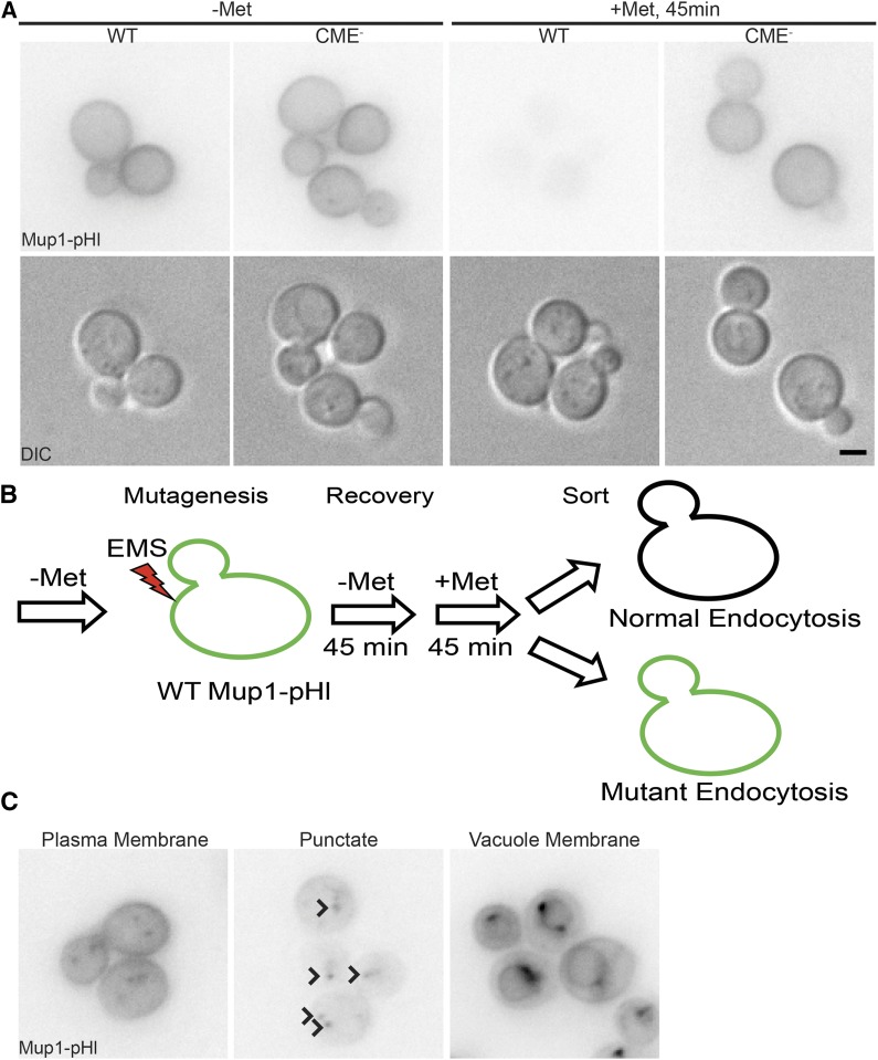
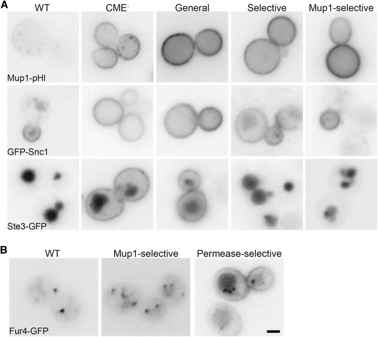
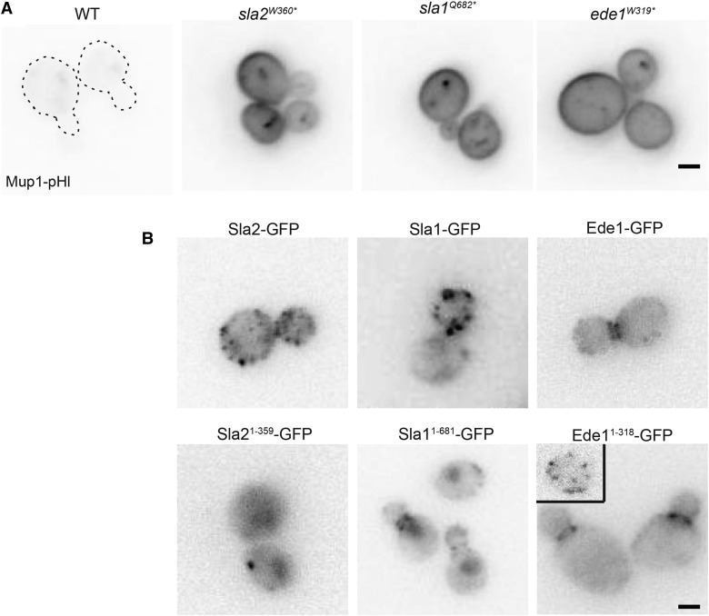
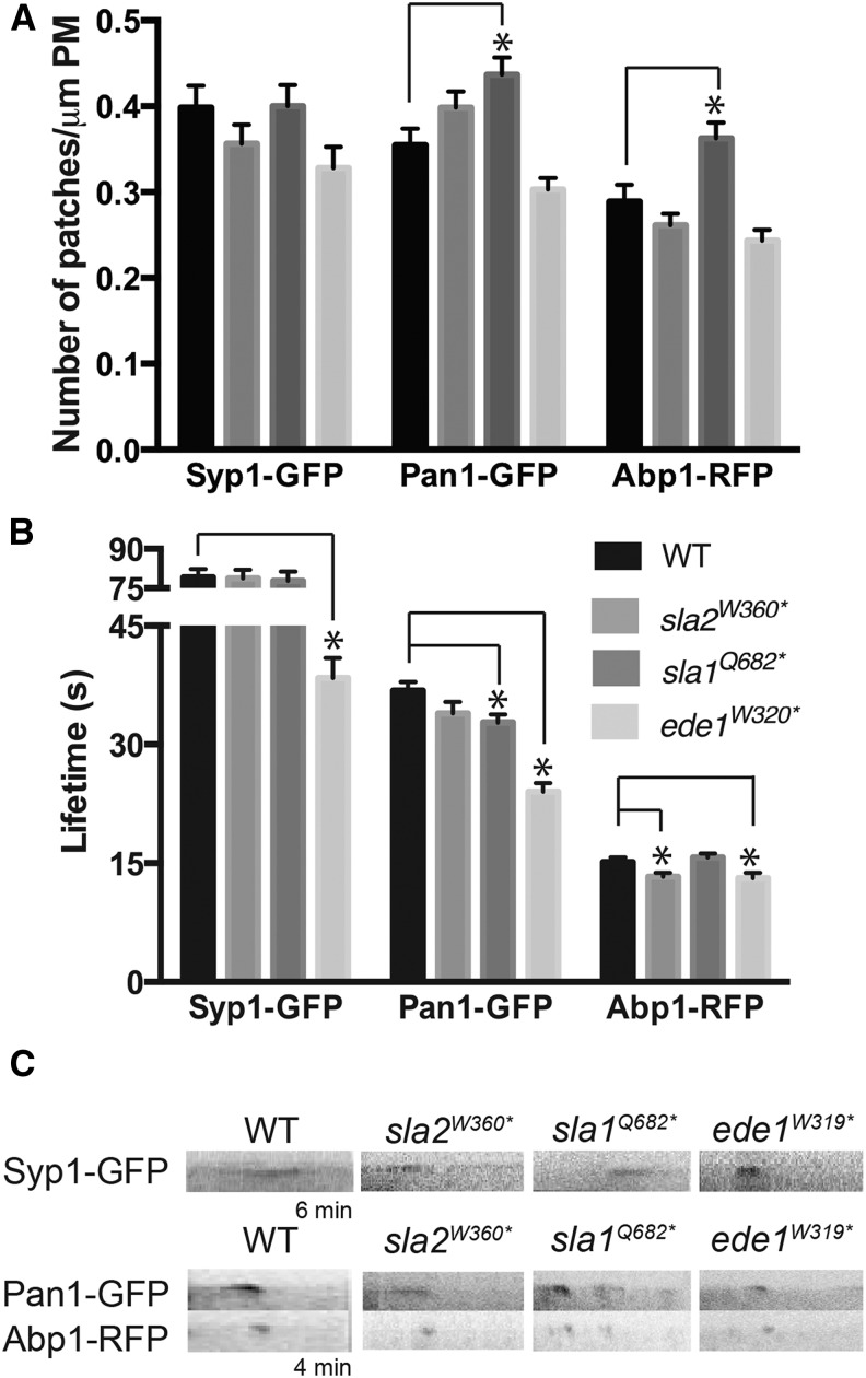
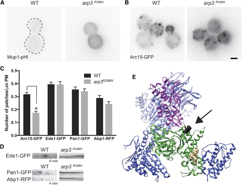
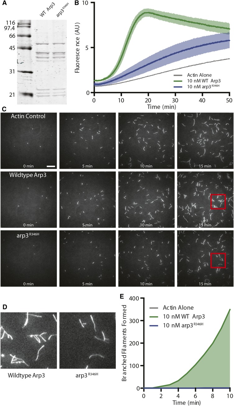
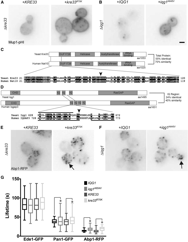
Endocytosis is a fundamental process for internalizing material from the plasma membrane, including many transmembrane proteins that are selectively internalized depending on environmental conditions. In most cells, the main route of entry is clathrin-mediated endocytosis (CME), a process that involves the coordinated activity of over 60 proteins; however, there are likely as-yet unidentified proteins involved in cargo selection and/or regulation of endocytosis. We performed a mutagenic screen to identify novel endocytic genes in Saccharomyces cerevisiae expressing the methionine permease Mup1 tagged with pHluorin (pHl), a pH-sensitive GFP variant whose fluorescence is quenched upon delivery to the acidic vacuole lumen. We used fluorescence-activated cell sorting to isolate mutagenized cells with elevated fluorescence, resulting from failure to traffic Mup1-pHl cargo to the vacuole, and further assessed subcellular localization of Mup1-pHl to characterize the endocytic defects in 256 mutants. A subset of mutant strains was classified as having general endocytic defects based on mislocalization of additional cargo proteins. Within this group, we identified mutations in four genes encoding proteins with known roles in endocytosis: the endocytic coat components SLA2, SLA1, and EDE1, and the ARP3 gene, whose product is involved in nucleating actin filaments to form branched networks. All four mutants demonstrated aberrant dynamics of the endocytic machinery at sites of CME; moreover, the arp3R346H mutation showed reduced actin nucleation activity in vitro Finally, whole genome sequencing of two general endocytic mutants identified mutations in conserved genes not previously implicated in endocytosis, KRE33 and IQG1, demonstrating that our screening approach can be used to identify new components involved in endocytosis.

Keywords: Arp2/3; actin; cargo sorting; clathrin-mediated endocytosis; forward genetic screen.


Figures

Figure 1
Figure 1
Endocytic function assayed during an EMS mutagenesis screen using an endogenous cargo, Mup1. A) Mup1-pHl localization was imaged using live-cell fluorescence microscopy (upper panel) and DIC optics (lower panel). WT and endocytic defective (CME-: ent1Δ ent2Δ yap1801Δ yap1802Δ +ENTH1) strains were imaged in medium lacking methionine (-Met) and 45 min after addition of 20 μg/ml of methionine (+Met) B) Schematic of mutagenesis screen workflow. C) Representative images of the three classes of mutants (Plasma membrane, Punctate, and Vacuole Membrane, as indicated) defined by Mup1-pHl localization 45 min after methionine addition. Arrowheads indicate punctae in the Punctate Class image. Scale bar, 2 μm.
Figure 2
Figure 2
Subclassification of the Plasma Membrane class mutants based on differential internalization of endocytic cargos. A) WT, CME-, and mutant strains representing general, selective, and Mup1-selective subclasses of mutants were imaged via fluorescence microscopy to observe differences in localization of fluorescently-tagged endocytic cargoes, Mup1-pHl (top row), GFP-Snc1 (middle row), and Ste3-GFP (bottom row). Mup1-pHl was imaged by live-cell microscopy after a 45 min methionine treatment. GFP-Snc1, and Ste3-GFP were expressed from low-copy plasmids. B) Fur4-GFP expressed in WT and mutant strains representing Mup1-selective and permease-selective subclasses were grown in YNB medium lacking uracil. 20 μg/ml of uracil was added for 1 h prior to to induce internalization of Fur4-GFP for imaging by live-cell microscopy. Scale bar, 2 μm.
Figure 3
Figure 3
SLA2, SLA1, and EDE1 truncations cause defects in endocytosis A) In a WT strain expressing Mup1-pHl, the specified amino acid in SLA2, SLA1, or EDE1 was mutated to a stop codon, generating the truncations. Mup1-pHl localization was assessed by fluorescence microscopy 45 min after addition of methionine. Dashed lines indicate the plasma membrane of WT Mup1-pHl cells. B) Strains expressing full-length and truncated Sla2, Sla1, and Ede1 were generated by the integration of GFP tag at the endogenous locus and imaged by fluorescence microscopy. Scale bar, 2μm.
Figure 4
Figure 4
Effect of SLA2, SLA1, and EDE1 truncation on endocytic patch number and lifetime of endocytic machinery proteins. A) Images of Syp1-GFP, Pan1-GFP and Abp1-RFP were acquired by live-cell time-lapse fluorescence microscopy, and the number of patches per micrometer of plasma membrane was counted for an equatorial section (N > 30 cells/strain). B) Lifetimes of Syp1-GFP, Pan1-GFP and Abp1-RFP calculated from movies of 120 frames acquired at intervals of 3 s (Syp1- GFP) or 2 s (Pan1-GFP, Abp1-RFP) (N > 50 patches/strain). All values from panels A and B are presented as mean ± SEM, * P < 0.05. C) Representative kymographs of Syp1-GFP, Pan1-GFP and Abp1-RFP in WT, sla2W360*, sla1Q682*and ede1W319* strains. Kymographs were made from movies used for the quantification, and are oriented with cell interior at the bottom.
Figure 5
Figure 5
A missense mutation in ARP3 causes endocytic defects A) In WT and arp3R346H expressing Mup1-pHl, cells were imaged by live cell microscopy 45 min after the addition of 20 μg/ml methionine. A dashed line indicates the plasma membrane of WT Mup1-pHl expressing cells. B) Images of Arc15-GFP in WT and arp3R346H cells were acquired by live-cell fluorescence microscopy. Scale bar, 2μm C) Time-lapse imaging of WT and arp3R346H cells expressing Arc15-GFP, Ede1-GFP, Pan1-GFP and Abp1-RFP were acquired, and the number of patches were per micrometer of plasma membrane was counted for an equatorial sections (N > 30 cells/strain). All values are presented as mean ± SEM, * P < 0.05. D) Representative kymographs of Ede1-GFP, Pan1-GFP and Abp1-RFP in WT, and arp3R346H strains. Kymographs were made from movies used for the quantification, and are oriented with cell interior at the bottom. E) Rendering was performed with Chimera (Pettersen et al. 2004) using the PDB file 3RSE of the bovine Arp2/3 structure (Ti et al. 2011). Arp3 is shown in green, Arp2 in purple, VCA in tan and the rest of the Arp2/3 complex members in blue. The amino acid in bovine Arp3 (R312) that corresponds to Arp3 (R346) in yeast shown in black.
Figure 6
Figure 6
Biochemical characterization of Arp2/3 complex vs. Arp2/Arp3R346H A) Side-by-side comparison of WT vs. Arp3R346H complex purified in vitro. B) Bulk pyrene-actin assembly assay comparing nucleation activity of Arp complex with WT vs. Arp3R346H with Las17 VCA. Data averaged from n = 3 independent experiments. C) Representative time-lapse images from TIRF experiments containing 0.5 μM actin (10% OG-labeled) and 10 nM Arp2/3 complex containing wildtype or arp3R346H as indicated. Experiments containing Arp2/3 complex also contain 100 nM GST-Las17 VCA to stimulate Arp2/3 activation. Scale bar, 20μm. D) Magnified view of the regions boxed in red, indicated in C. E) Quantification of the number of branched filaments nucleated in the same reactions as D. Data combined from 6 fields of view per experiment, with 2-3 independent experiments (12-18 fields of view, total). Note, at 7 min arp3R346H nucleated a single, persistent branched actin filament. All values are presented as mean ± SEM.
Figure 7
Figure 7
Effect of kre33R70K and iqg1A654V mutations on Mup1-pHl internalization A) In kre33Δ cells expressing Mup1-pHl, either KRE33 or kre33R70K was expressed from a low-copy plasmid. B) Either IQG1 or iqg1A654V was expressed from a low copy plasmid in an iqg1Δ strain expressing Mup1-pHl. Mup1-pHl images were acquired 45 min after addition of 20 μg/ml methionine. Scale bar, 2μm. C) Schematics of Saccharomyces cerevisiae (Yeast) Kre33 and Human homolog Nat10 protein domains showing an alignment surrounding the mutation (black arrow) using Pfam and Sequence Manipulation Suite (Stothard 2000; Finn et al. 2016) D) Schematics of Saccharomyces cerevisiae (Yeast) Iqg1 and Human homolog IQGAP3 protein domains showing an alignment surrounding the mutation (black arrow) using Pfam and Sequence Manipulation Suite. Amino acids included within IQ motifs are indicated with black bars; below for Human IQGAP3 and above for Yeast Iqg1. E) Either KRE33 or kre33R70K was expressed from a low-copy plasmid in kre33Δ cells expressing Abp1-RFP. Arrow indicates aberrant actin patches. F) In an iqg1Δ strain expressing Abp1-RFP, either IQG1 or iqg1A654V was expressed from a low copy plasmid. Arrows indicate aberrant actin patches. G) Lifetimes of Ede1-GFP, Pan1-GFP and Abp1-RFP calculated from movies of 120 frames acquired at intervals of 3 s (Syp1- GFP) or 2 s (Pan1-GFP, Abp1-RFP) (N > 50 patches/strain). The central lines of the box plot correspond to the median and the whiskers denote the min and max lifetimes. * P < 0.05 using an F test.

Similar articles

Cited by

References

    1. Aghamohammadzadeh S., Ayscough K. R., 2009. Differential requirements for actin during yeast and mammalian endocytosis. Nature Publishing Group 11: 1039–1042. 10.1038/ncb1918 - DOI - PMC - PubMed
    1. Aguilar R. C., Longhi S. A., Shaw J. D., Yeh L.-Y., Kim S., et al. , 2006. Epsin N-terminal homology domains perform an essential function regulating Cdc42 through binding Cdc42 GTPase-activating proteins. Proc. Natl. Acad. Sci. USA 103(11): 4116–4121. 10.1073/pnas.0510513103 - DOI - PMC - PubMed
    1. Ayscough K. R., Stryker J., Pokala N., Sanders M., Crews P., et al. , 1997. High rates of actin filament turnover in budding yeast and roles for actin in establishment and maintenance of cell polarity revealed using the actin inhibitor latrunculin-A. J. Cell Biol. 137(2): 399–416. 10.1083/jcb.137.2.399 - DOI - PMC - PubMed
    1. Barker S. L., Lee L., Pierce B. D., Maldonado-Baez L., Drubin D. G., et al. , 2007. Interaction of the endocytic scaffold protein Pan1 with the type I myosins contributes to the late stages of endocytosis. Mol. Biol. Cell 18(8): 2893–2903. 10.1091/mbc.E07-05-0436 - DOI - PMC - PubMed
    1. Bitsikas V., Corrêa I. R., Nichols B. J., 2014. Clathrin-independent pathways do not contribute significantly to endocytic flux. eLife 3: e03970 10.7554/eLife.03970 - DOI - PMC - PubMed

Publication types

MeSH terms

LinkOut - more resources