. 2018 May 4;293(18):6844-6858.
doi: 10.1074/jbc.RA117.000164. Epub 2018 Mar 14.

An activating mutation of interferon regulatory factor 4 (IRF4) in adult T-cell leukemia

Affiliations

An activating mutation of interferon regulatory factor 4 (IRF4) in adult T-cell leukemia

Mathew A Cherian et al. J Biol Chem. .

Abstract

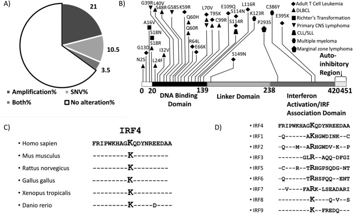
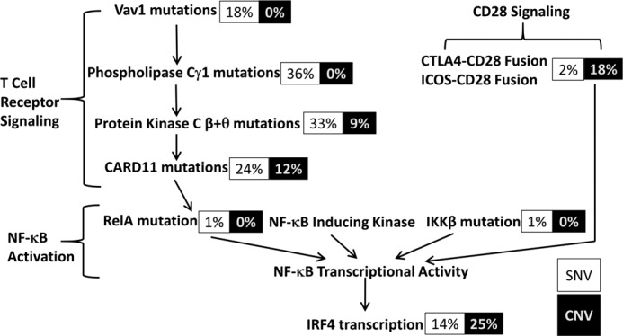
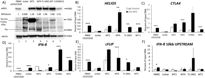
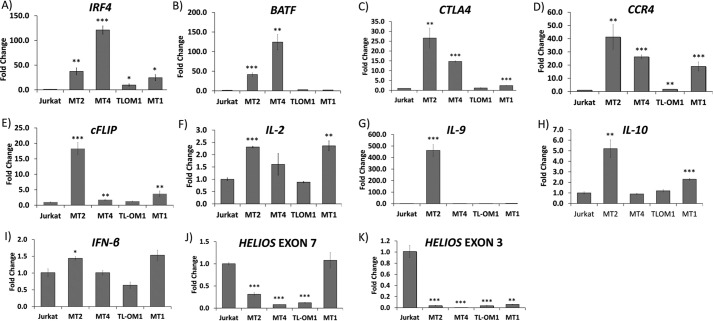
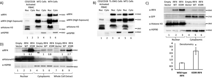
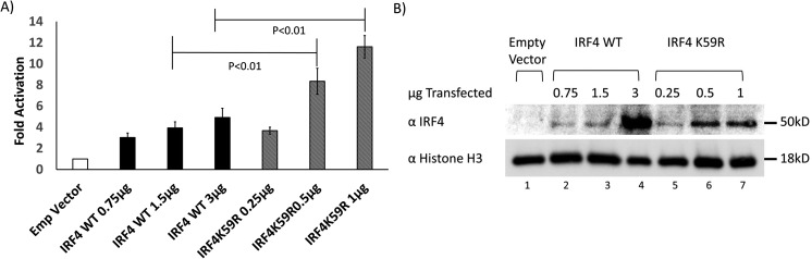
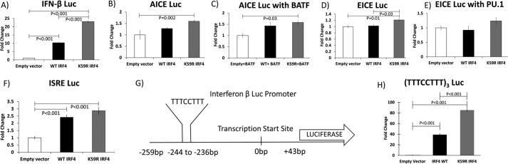
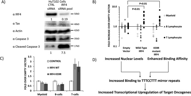
The human T-cell leukemia virus-1 (HTLV-1) oncoprotein Tax drives cell proliferation and resistance to apoptosis early in the pathogenesis of adult T-cell leukemia (ATL). Subsequently, probably as a result of specific immunoediting, Tax expression is down-regulated and functionally replaced by somatic driver mutations of the host genome. Both amplification and point mutations of interferon regulatory factor 4 (IRF4) have been previously detected in ATL., K59R is the most common single-nucleotide variation of IRF4 and is found exclusively in ATL. High-throughput whole-exome sequencing revealed recurrent activating genetic alterations in the T-cell receptor, CD28, and NF-κB pathways. We found that IRF4, which is transcriptionally activated downstream of these pathways, is frequently mutated in ATL. IRF4 RNA, protein, and IRF4 transcriptional targets are uniformly elevated in HTLV-1-transformed cells and ATL cell lines, and IRF4 was bound to genomic regulatory DNA of many of these transcriptional targets in HTLV-1-transformed cell lines. We further noted that the K59R IRF4 mutant is expressed at higher levels in the nucleus than WT IRF4 and is transcriptionally more active. Expression of both WT and the K59R mutant of IRF4 from a constitutive promoter in retrovirally transduced murine bone marrow cells increased the abundance of T lymphocytes but not myeloid cells or B lymphocytes in mice. IRF4 may represent a therapeutic target in ATL because ATL cells select for a mutant of IRF4 with higher nuclear expression and transcriptional activity, and overexpression of IRF4 induces the expansion of T lymphocytes in vivo.

Keywords: ATL; HTLV; NF-kappaB transcription factor; cancer biology; chromatin immunoprecipitation (ChiP); driver mutation; interferon regulatory factor (IRF); retrovirus.


Conflict of interest statement

The authors declare that they have no conflicts of interest with the contents of this article

Figures

Figure 1.
Figure 1.
IRF4 alterations in ATL. A, pie chart depicting types of genetic alterations of IRF4 in ATL. B, distribution of SNVs across the IRF4 protein sequence in lymphoid malignancies, including ATL, obtained from the COSMIC database shows a predominance of SNVs in the DNA-binding domain. K59R and L70V, the most common recurrent mutations in ATL, are located in the DNA-binding domain. C, the Lys-59 region of IRF4 is conserved across species. D, sequences of other IRFs incorporate a lysine or arginine at the position corresponding to IRF4 Lys-59.
Figure 2.
Figure 2.
IRF4 lies downstream of constitutively activated T-cell receptor and CD28 pathways in ATL cells. Shown are the percentage of cases in which each of the proteins upstream of IRF4 in the T-cell receptor, CD28, and NF-κB pathways is mutated in Kataoka et al. (22). SNVs are depicted in black type and copy number variations (CNV) in white type. Note that in ATL, protein kinase Cβ is exclusively altered by point mutation and protein kinase Cθ exclusively by amplification.
Figure 3.
Figure 3.
IRF4 is overexpressed in ATL and bound to transcriptional target genes. A, Western blotting demonstrating IRF4 expression in CD3/CD28-activated PBMCs and Jurkat, MT2, MT4, TL-OM1, MT1, and ED40515 cell lines. An immunoblot shows Tax protein expression. Env-Tax fusion protein in lane 3 of the Tax blot (middle) is seen between 50 and 75 kDa and indicated by the top arrow. Tax-specific bands are seen in lanes 3 and 4 of the Tax blot at 40 kDa, as indicated by the bottom arrow. Immunoblot for actin (bottom) shows actin expression as loading control. B–F, binding of IRF4 to HELIOS 3′-regulatory region (B), CTLA4 gene (C), IFNβ gene (D), cFLIP gene (E), and a control genomic region 10 kilobases upstream of the IRF4 start site (F) by IRF4, as demonstrated by a ChIP assay in CD3/CD28-activated PBMCs and Jurkat, MT2, MT4, TL-OM1, and MT1 cells. B–F, results of four replicates. Error bars, S.E. *, p < 0.05; **, p < 0.01; ***, p < 0.001.
Figure 4.
Figure 4.
Expression of IRF4 and IRF4-target genes in HTLV-1–transformed and ATL cell lines. A–K, RNA expression of IRF4 and its transcriptional targets by qRT-PCR: IRF4 (A), BATF (B), CTLA4 (C), CCR4 (D), c-FLIP (E), IL-2 (F), IL-9 (G), IL-10 (H), IFN-β (I), HELIOS exon 7 (J), and exon 3 (K) in Jurkat, MT2, MT4, TL-OM1, and MT1 cells. Error bars (representing S.E.) are indicated as well as p values for comparisons with Jurkat; *, p < 0.05; **, p < 0.01; ***, p < 0.001. The values represent average of 3 replicates.
Figure 5.
Figure 5.
Preferential nuclear localization of WT and K59R IRF4. A, Western blotting of nuclear and cytoplasmic fractions of IRF4 in CD3/CD28 activated PBMCs and MT2 and MT4 cells showing predominantly nuclear localization of IRF4. B, Western blotting of nuclear and cytoplasmic fractions of IRF4 in CD3/CD28-activated PBMCs and TL-OM1 and MT1 cells, demonstrating predominantly nuclear localization of IRF4. C, Western blotting depicting expression of IRF4 in nuclear and cytoplasmic fractions of 293T cells transiently transfected with equal mass of MSCV-Empty-IRES-GFP, MSCV-IRF4 WT-IRES-GFP, and MSCV-IRF4 K59R-IRES-GFP. Lanes 1–3 show nuclear fractions. Lanes 4–6 show cytosolic fractions. The IRF4 immunoblot shows increased nuclear levels of the K59R mutant of IRF4 (top blot, lane 3) as compared with WT IRF4 (lane 2). Lanes 5 and 6 show equal IRF4 expression in the cytosolic fraction. The GFP blot shows equal expression of IRF4 in the cytosolic fraction from the IRES sequence of the vectors, indicating equal transfection efficiency. The HSP90 blot shows equal loading of cytosolic fractions, and lack of signal in the nuclear fractions confirms the purity of the subcellular fractions. The histone H3 blot shows equal loading of nuclear fractions, and lack of signal in the cytosolic fraction further confirms the purity of the subcellular fractions. A bar graph depicts results from three replicates of the experiment depicted in C. *, p < 0.05; **, p < 0.01; ***, p < 0.001. D, Western blotting depicting expression of IRF4 in nuclear and cytoplasmic fractions of Jurkat T cells transiently transfected with equal mass of MSCV-Empty, MSCV-IRF4 WT, and MSCV-IRF4 K59R. Lanes 1–3, nuclear fractions; lanes 4–6, cytosolic fractions; lanes 7–9, whole-cell lysates. The IRF4 immunoblot shows increased nuclear levels of the K59R mutant of IRF4 (top blot, lane 3) as compared with WT IRF4 (lane 2). Lanes 5 and 6 show equal IRF4 expression in the cytosolic fraction. The HSP90 blot shows equal loading of cytosolic fractions, and lack of signal in the nuclear fractions confirms the purity of the subcellular fractions. The histone H3 blot shows equal loading of the nuclear fractions, and lack of signal in the cytosolic fraction further confirms the purity of the subcellular fractions.
Figure 6.
Figure 6.
Effects of WT and K59R IRF4 on interferon β promoter. A, luciferase expression from PGL2 vector with proximal promoter region of interferon β expressed as a ratio to Renilla Luc expressed from the constitutive thymidine kinase promoter in 293T cells, co-transfected with empty vector or increasing mass of MSCV-IRF4 WT-IRES-GFP plasmid or MSCV-IRF4 K59R-IRES-GFP plasmid; MSCV-IRF4 WT plasmid was transfected at a 3:1 ratio to MSCV-IRF4 K59R-IRES-GFP plasmid to equalize nuclear expression levels while keeping total DNA constant. 3 μg of total DNA was transfected (3 μg of empty vector DNA in bar 1 and the amount of empty vector decreased as IRF4 vector was increased to keep a total of 3 μg. The luciferase graph represents results of five replicates. Error bars, S.E. Significance was calculated using unpaired Student's t test. B, Western blotting showing expression of IRF4 in nuclear lysates corresponding to samples for the experiment shown in A.
Figure 7.
Figure 7.
Effects of WT and K59R IRF4 on transcriptional reporter elements in 293T cells. A, -fold induction of firefly luciferase expressed from the interferon β proximal promoter by WT S-tagged IRF4 and S-tagged K59R mutant IRF4 over empty vector expressed as a ratio to Renilla luciferase expressed from thymidine kinase promoter. B, induction of firefly luciferase expressed from an AP1-IRF composite element by S-tagged WT IRF4 and S-tagged K59R mutant IRF4 expressed as a ratio to Renilla luciferase expressed from thymidine kinase promoter. C, induction of firefly luciferase expressed from an AP1-IRF composite element by S-tagged WT IRF4 and S-tagged K59R mutant IRF4 transfected in combination with BATF expressed as a ratio to Renilla luciferase expressed from thymidine kinase promoter. D, induction of firefly luciferase expressed from an Ets-IRF composite element by S-tagged WT IRF4 and S-tagged K59R mutant IRF4 expressed as a ratio to Renilla luciferase expressed from thymidine kinase promoter. E, induction of firefly luciferase expressed from an Ets-IRF composite element by S-tagged WT IRF4 and S-tagged K59R mutant IRF4 in combination with PU.1 expressed as a ratio to Renilla luciferase expressed from thymidine kinase promoter. F, induction of firefly luciferase expressed from an ISRE element by WT IRF4 and K59R mutant IRF4 expressed as a ratio to Renilla luciferase expressed from thymidine kinase promoter. G, sequence from the IFN-β Luc plasmid corresponding to nucleotides −259 to +43 relative to the transcription start site was sequenced from the interferon β Luc plasmid used in experiments depicted in Figs. 6A and 7A. A mirror repeat element in that sequence is highlighted. H, induction of firefly luciferase expressed from a mirror repeat element by S-tagged WT IRF4 and S-tagged K59R mutant IRF4 expressed as a ratio to Renilla luciferase expressed from thymidine kinase promoter. A–G, results of four replicates; H, results of three replicates.
Figure 8.
Figure 8.
Effect of WT and K59R IRF4 on T-lymphocyte abundance in mice. A, Western blotting depicting expression of cleaved caspase-3 (as a measure of apoptosis), total caspase-3, IRF4, Tax, and actin in lysates of HuT102 cells treated with control siRNA or a pool of siRNAs to IRF4. B and C, murine hematopoietic stem cells (CD45.1, c-kit+, Sca-1+ bone marrow cells) were transduced with MSCV retroviral vectors, which express IRF4, IRF4 (K59R), or vector alone, as well as GFP from an IRES and engrafted into irradiated CD45.2 recipients (n = 15, 10, and 10 for control, WT IRF4–overexpressing, and K59R-overexpressing mice). Four weeks after engraftment, peripheral blood was collected, and FACS was used to determine the abundance of CD45.1, GFP+ myeloid cells (Gr-1+, Mac-1+), B cells (B220+), and T cells (CD4+, CD8+). Whereas no effect was observed on B cells or myeloid cells, the T-cell populations in IRF4-overexpressing mice were significantly greater than controls, demonstrating that overexpression of IRF4 or IRF4 (K59R) results in T-cell proliferation in vivo. D, mechanisms for increased oncogenic activity of the ATL-specific IRF4 K59R mutation.

Similar articles

Cited by

References

    1. Uchiyama T., Yodoi J., Sagawa K., Takatsuki K., and Uchino H. (1977) Adult T-cell leukemia: clinical and hematologic features of 16 cases. Blood 50, 481–492 - PubMed
    1. Tamura K. (1996) [Clinical classification of adult T-cell leukemia and its complications]. Rinsho Byori 44, 19–23 - PubMed
    1. Shimoyama M. (1991) Diagnostic criteria and classification of clinical subtypes of adult T-cell leukaemia-lymphoma: a report from the Lymphoma Study Group (1984–87). Br. J. Haematol. 79, 428–437 10.1111/j.1365-2141.1991.tb08051.x - DOI - PubMed
    1. Teshima T., Akashi K., Shibuya T., Taniguchi S., Okamura T., Harada M., Sumida I., Hanada M., and Niho Y. (1990) Central nervous system involvement in adult T-cell leukemia/lymphoma. Cancer 65, 327–332 10.1002/1097-0142(19900115)65:2%3C327::AID-CNCR2820650224%3E3.0.CO%3B2-W - DOI - PubMed
    1. Ma W. L., Li C. C., Yu S. C., and Tien H. F. (2014) Adult T-cell lymphoma/leukemia presenting as isolated central nervous system T-cell lymphoma. Case Rep. Hematol. 2014, 917369 - PMC - PubMed

Publication types

MeSH terms