. 2018 May 4;293(18):6905-6914.
doi: 10.1074/jbc.RA117.001670. Epub 2018 Mar 14.

Structural and biochemical characterization of Siw14: A protein-tyrosine phosphatase fold that metabolizes inositol pyrophosphates

Affiliations

Structural and biochemical characterization of Siw14: A protein-tyrosine phosphatase fold that metabolizes inositol pyrophosphates

Huanchen Wang et al. J Biol Chem. .

Abstract

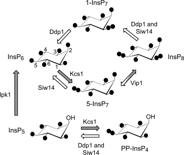
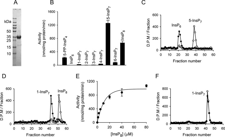
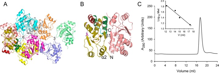
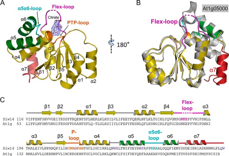
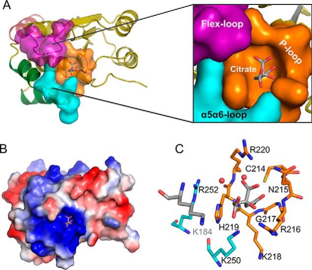
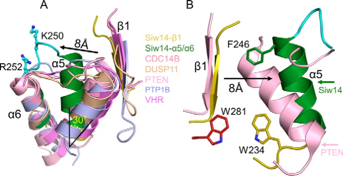
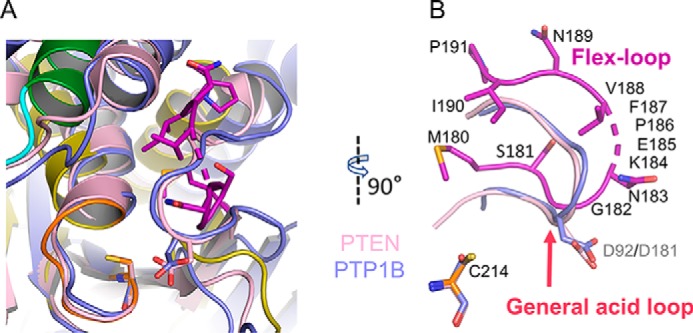
Inositol pyrophosphates (PP-InsPs) are "energetic" intracellular signals that are ubiquitous in animals, plants, and fungi; structural and biochemical characterization of PP-InsP metabolic enzymes provides insight into their evolution, reaction mechanisms, and regulation. Here, we describe the 2.35-Å-resolution structure of the catalytic core of Siw14, a 5-PP-InsP phosphatase from Saccharomyces cerevisiae and a member of the protein tyrosine-phosphatase (PTP) superfamily. Conclusions that we derive from structural data are supported by extensive site-directed mutagenesis and kinetic analyses, thereby attributing new functional significance to several key residues. We demonstrate the high activity and exquisite specificity of Siw14 for the 5-diphosphate group of PP-InsPs. The three structural elements that demarcate a 9.2-Å-deep substrate-binding pocket each have spatial equivalents in PTPs, but we identify how these are specialized for Siw14 to bind and hydrolyze the intensely negatively charged PP-InsPs. (a) The catalytic P-loop with the CX5R(S/T) PTP motif contains additional, positively charged residues. (b) A loop between the α5 and α6 helices, corresponding to the Q-loop in PTPs, contains a lysine and an arginine that extend into the catalytic pocket due to displacement of the α5 helix orientation through intramolecular crowding caused by three bulky, hydrophobic residues. (c) The general-acid loop in PTPs is replaced in Siw14 with a flexible loop that does not use an aspartate or glutamate as a general acid. We propose that an acidic residue is not required for phosphoanhydride hydrolysis.

Keywords: crystal structure; dual-specificity phosphoprotein phosphatase; enzyme mechanism; inositol phosphate; phosphatase.


Conflict of interest statement

The authors declare that they have no conflicts of interest with the contents of this article

Figures

Figure 1.
Figure 1.
Inositol pyrophosphate metabolism in S. cerevisiae. A proposed cyclical pathway of PP-InsP turnover (see Ref. 1) is shown. Filled circles represent phosphate groups; their locants are given for InsP6, and these apply to the other molecules as each is oriented identically. The arrows depicting the various reactions are colored white to denote phosphatase activity and gray to describe kinase activity. Enzyme nomenclature is as follows: Ddp1, diphosphoinositol-polyphosphate phosphatase; Ipk1, inositol-pentakisphosphate kinase; Kcs1, inositol-hexakisphosphate kinase; Vip1, diphosphoinositol-pentakisphosphate kinase.
Figure 2.
Figure 2.
Catalytic activities of recombinant Siw14. A, SDS-PAGE of purified full-length Siw14. B, substrate specificity of Siw14. Each of the inositol phosphates was present at a concentration of 10 μm and incubated with 0.09–1.8 μg of Siw14 for 30 min. Vertical bars are means with S.E. (error bars) obtained from three to six experiments. C, representative HPLC analysis of assays containing 120 ng of Siw14 incubated with 5-[3H]InsP7 for 15 min (closed circles) plus a zero-time control (open circles). The elution peaks of the 5-[3H]InsP7 substrate and [3H]InsP6 product are observed in fraction numbers 38 and 21, respectively. D, representative HPLC analysis of assays containing 120 ng of Siw14 incubated with 1,5-[3H]InsP8 for 15 min (closed circles) plus a zero-time control (open circles). The peak elution time for the [3H]InsP7 product is observed in fraction number 42, and hence it is identified as 1-[3H]InsP7 (see F). E, substrate-saturation plot for Siw14-mediated dephosphorylation of 1,5-InsP8; data are means with S.E. (error bars) from five experiments. F, representative HPLC analysis of assays containing 120 ng of Siw14 incubated with 1-[3H]InsP7 for 15 min (closed circles) plus a zero-time control (open circles). The elution peak of 1-[3H]InsP7 is fraction number 42.
Figure 3.
Figure 3.
Crystal packing of Siw14. A, shown are the interfaces of the trimer (denoted as 3) and hexamer (denoted as 6). Ribbon structures are shown with various colors for different chains. B, the dimer interface and structural elements involved are denoted. C, analysis of recombinant Siw14(116–281) purified by gel filtration. Mwt, molecular weight.
Figure 4.
Figure 4.
Crystal structure of Siw14. A, overall structure of Siw14(116–281). A citrate molecule was observed in the structure. The 2FoFc electron density map was contoured at 1.0 σ. B, superimposition of Siw14 (in color) and At1g05000 (in gray; Protein Data Bank code 1XRI) catalytic domains; core root mean square deviation is 0.654 Å. C, alignment of residues 116–281 of Siw14 (KZV08605) with corresponding residues from At1g05000 (At1g; AAO63274). Those residues that were targeted for site-directed mutagenesis are highlighted in color. The secondary structural elements of Siw14 are also shown above the alignment and are color-coded to match A and B.
Figure 5.
Figure 5.
The architecture of the Siw14 active site. A, surface representation of the substrate-binding pocket with a zoom-out to show its position in the overall protein structure. The P-loop is colored orange, the α5-α6 loop is colored cyan, and the flex-loop is colored purple (omitting residues 184–187, which have low electron density in the structure). Other structure elements are shown in ribbon. B, electrostatic surface plot with blue and red coloration to denote positive and negative electrostatic potentials, respectively, at physiological pH; the captured citrate molecule is also shown. C, residues that surround the citrate are shown as sticks. Nitrogen is blue, sulfur is yellow, oxygen is red, and carbons are color-coded to indicate their parent structural feature (A). The two red spheres represent water molecules.
Figure 6.
Figure 6.
Orientation of the α5-α6 loop of Siw14 reflects intramolecular crowding. A, overlap of Siw14 β1 strand (shown in yellow) and α5 and α6 helices (shown in green) with class I PTPs, including CDC14B (Protein Data Bank code 1OHE; shown in dark pink), Dusp11 (Protein Data Bank code 4NYH; shown in wheat), PTEN (Protein Data Bank code 1D5R; shown in light pink), PTP1B (Protein Data Bank code 1PTY; shown in light blue), and VHR (Vaccinia H1-related phosphatase; Protein Data Bank code 1VHR; shown in light purple). Candidate substrate-binding residues Lys-250 and Arg-252 are shown in cyan with stick and ball modeling. We have highlighted the displacement of α5 relative to the corresponding α-helix in the other PTPs (by 30° and 8 Å). B, comparison of equivalent structural elements in both PTEN and Siw14. We have labeled the bulky, aromatic residues Phe-246, Trp-234, and Trp-281, which appear to contribute to the intramolecular crowding that underlies the displacement of the α5 helix. For Siw14, see Fig. 4 for the positions in the overall protein structure of the α5 and α6 helices and the β1 loop.
Figure 7.
Figure 7.
Absence of a general acid in Siw14. A, the catalytic center of Siw14 (green, α6 helix; orange stick, P-loop; purple stick, flex-loop) is superimposed upon PTEN (PDB code 1D5R; shown in light pink) and PTP1B (PDB code 1PTY; shown in light blue). B, highlight from A of the relative positions of the flex-loop of Siw14 compared with the general-acid loops of PTEN and PTP1B. Also shown, for the purposes of orientation, are each protein's catalytic cysteine residues, although in this perspective they cannot be individually distinguished; Cys-214 refers to Siw14. The numbers in gray font are the general-acid residues Asp-92 for PTEN and Asp-181 for PTP1B. The numbers in black font correspond to residues in Siw14; broken stick indicates the four residues that are disordered in our structure.

Similar articles

Cited by

References

    1. Shears S. B. (2018) Intimate connections: inositol pyrophosphates at the interface of metabolic regulation and cell signaling. J. Cell. Physiol. 233, 1897–1912 10.1002/jcp.26017 - DOI - PMC - PubMed
    1. Thota S. G., and Bhandari R. (2015) The emerging roles of inositol pyrophosphates in eukaryotic cell physiology. J. Biosci. 40, 593–605 10.1007/s12038-015-9549-x - DOI - PubMed
    1. Azevedo C., and Saiardi A. (2017) Eukaryotic phosphate homeostasis: the inositol pyrophosphate perspective. Trends Biochem. Sci. 42, 219–231 10.1016/j.tibs.2016.10.008 - DOI - PubMed
    1. Chakraborty A. (2017) The inositol pyrophosphate pathway in health and diseases. Biol. Rev. Camb. Philos. Soc. 10.1111/brv.12392 10.1111/brv.12392 - DOI - DOI - PMC - PubMed
    1. Saiardi A. (2016) Protein pyrophosphorylation: moving forward. Biochem. J. 473, 3765–3768 10.1042/BCJ20160710C - DOI - PMC - PubMed

Publication types

MeSH terms

Substances