. 2018 Apr 27;293(17):6517-6529.
doi: 10.1074/jbc.RA117.001073. Epub 2018 Mar 14.

The cellular stress proteins CHCHD10 and MNRR1 (CHCHD2): Partners in mitochondrial and nuclear function and dysfunction

Affiliations

The cellular stress proteins CHCHD10 and MNRR1 (CHCHD2): Partners in mitochondrial and nuclear function and dysfunction

Neeraja Purandare et al. J Biol Chem. .

Abstract

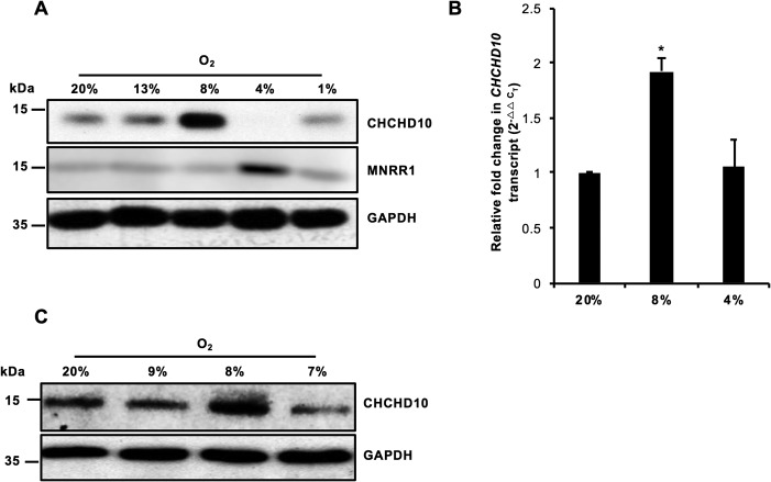
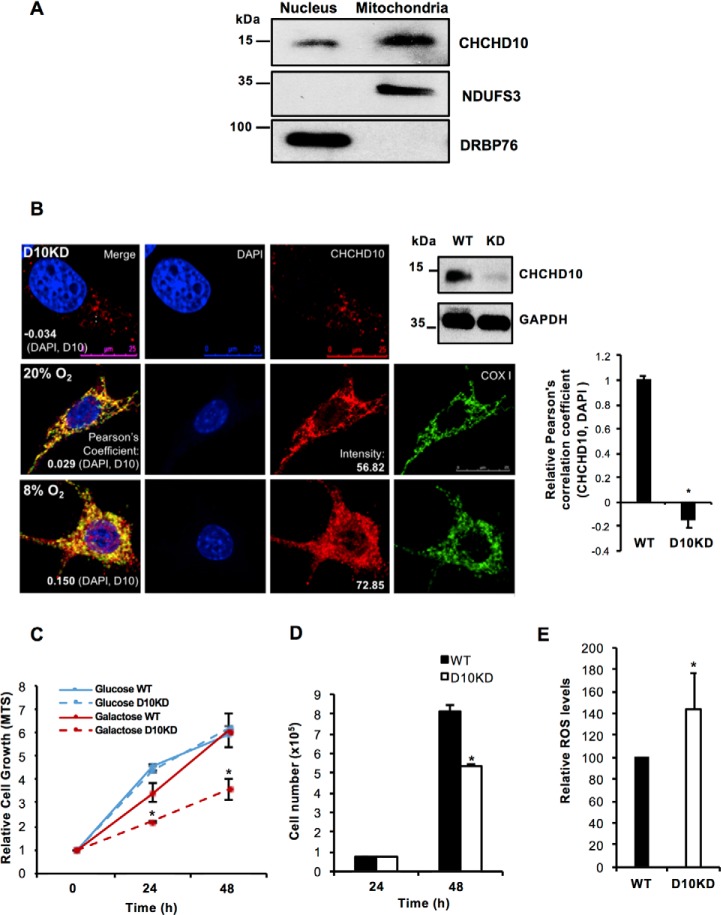
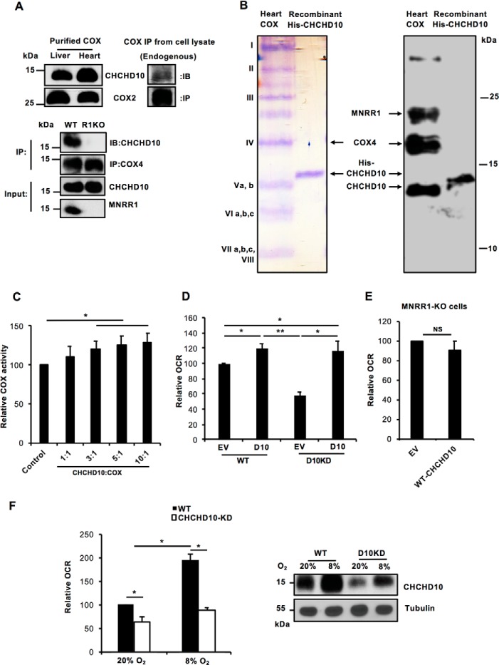
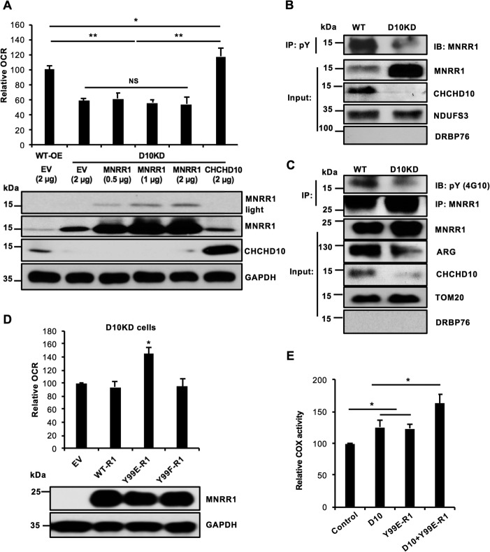
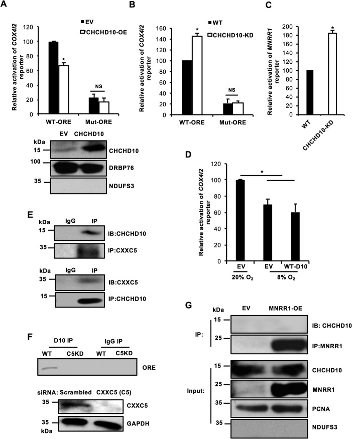
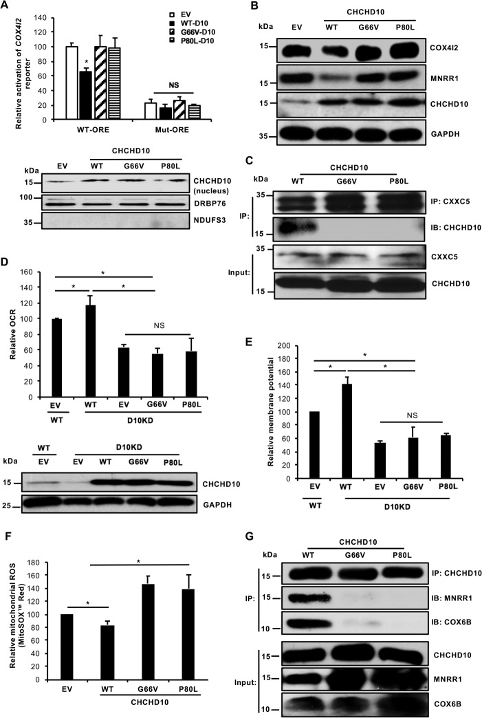
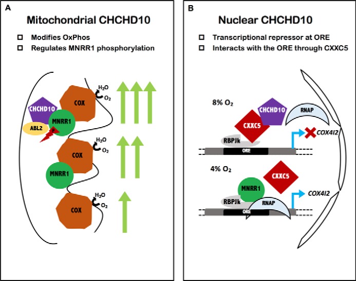
Coiled-coil-helix-coiled-coil-helix domain-containing 10 (CHCHD10) and CHCHD2 (MNRR1) are homologous proteins with 58% sequence identity and belong to the twin CX9C family of proteins that mediate cellular stress responses. Despite the identification of several neurodegeneration-associated mutations in the CHCHD10 gene, few studies have assessed its physiological role. Here, we investigated CHCHD10's function as a regulator of oxidative phosphorylation in the mitochondria and the nucleus. We show that CHCHD10 copurifies with cytochrome c oxidase (COX) and up-regulates COX activity by serving as a scaffolding protein required for MNRR1 phosphorylation, mediated by ARG (ABL proto-oncogene 2, nonreceptor tyrosine kinase (ABL2)). The CHCHD10 gene was maximally transcribed in cultured cells at 8% oxygen, unlike MNRR1, which was maximally expressed at 4%, suggesting a fine-tuned oxygen-sensing system that adapts to the varying oxygen concentrations in the human body under physiological conditions. We show that nuclear CHCHD10 protein down-regulates the expression of genes harboring the oxygen-responsive element (ORE) in their promoters by interacting with and augmenting the activity of the largely uncharacterized transcriptional repressor CXXC finger protein 5 (CXXC5). We further show that two genetic CHCHD10 disease variants, G66V and P80L, in the mitochondria exhibit faulty interactions with MNRR1 and COX, reducing respiration and increasing reactive oxygen species (ROS), and in the nucleus abrogating transcriptional repression of ORE-containing genes. Our results reveal that CHCHD10 positively regulates mitochondrial respiration and contributes to transcriptional repression of ORE-containing genes in the nucleus, and that genetic CHCHD10 variants are impaired in these activities.

Keywords: cell stress; cytochrome c oxidase (Complex IV); energy metabolism; hypoxia; mitochondria; mitochondrial disease; neurodegenerative disease; scaffolding protein; transcriptional regulator.


Conflict of interest statement

The authors declare that they have no conflicts of interest with the contents of this article

Figures

Figure 1.
Figure 1.
CHCHD10 is a hypoxia-sensitive gene. A, HeLa cells were incubated for 48 h at the stated O2 levels. Whole cell lysates were separated on an SDS-PAGE gel and analyzed for CHCHD10 and MNRR1 levels. GAPDH was probed as a loading control. B, real-time PCR analysis of transcript levels of endogenous CHCHD10 at 20, 8, and 4% oxygen (n = 4; *, p < 0.05). C, as in A but not probed for MNRR1. Error bars in graphs represent 1 standard deviation from the mean of repeat determinations.
Figure 2.
Figure 2.
CHCHD10 is localized to both the nucleus and the mitochondria. A, nuclear and mitochondrial fractions (HEK293 cells) were analyzed for levels of CHCHD10. Effectiveness of fractionation was shown using DRBP76 as a nuclear marker and NDUFS3 as a mitochondrial marker. B, representative images of CHCHD10 (red) colocalization with the nucleus (DAPI, blue; merge, purple) in HEK293 WT and CHCHD10-KD cells. The Pearson correlation coefficient values (negative in the KD cells) indicate the specificity of nuclear signal of CHCHD10 (top panel). To show effectiveness of CHCHD10 knockdown, equal amounts of lysate from HEK293 WT and CHCHD10-KD cells were probed for CHCHD10 levels (top panel, right). GAPDH was probed as loading control. Representative images of CHCHD10 (red) localization in HEK293 cells at normoxia (20% O2) (middle panel) and hypoxia (8% O2) (bottom panel). Right, relative Pearson correlation coefficient values (negative in the KD cells) indicate the specificity of nuclear signal of CHCHD10 (n = 4; *, p < 0.05). C, equal numbers of HEK293 WT and CHCHD10-KD cells were plated and grown in medium containing either glucose or galactose as the primary carbon source. Growth was assessed in real time using CellTiter 96 AQueous One Solution Cell Proliferation Assay (MTS) (n = 4; *, p < 0.05 for CHCHD10-KD as compared with WT grown in galactose; NS for glucose). D, equal numbers of HEK293 WT and CHCHD10-KD cells were plated and grown in galactose medium (n = 4 each in quadruplicate; *, p < 0.05). E, total cellular ROS was measured from WT and CHCHD10-KD HEK293 cells using CM-H2DCFDA (n = 4; *, p < 0.05). NS, not significant. Error bars in graphs represent 1 standard deviation from the mean of repeat determinations.
Figure 3.
Figure 3.
CHCHD10 regulates COX activity in the mitochondria via phosphorylation of MNRR1. A, upper, purified COX from cow liver and heart and mitochondrial fractions from HEK293 cells were probed for CHCHD10. COX2 is probed as a loading control for the tissue COX, and COX2 beads were used to immunoprecipitate endogenous COX from HEK293 cells. All samples were probed for CHCHD10. Lower, to show lack of COX binding of CHCHD10 in MNRR1-KO cells, both WT and HEK293 MNRR1-KO cells were transfected with an expression plasmid for WT-CHCHD10 (FLAG-tagged). After 48 h, COX was immunoprecipitated from mitochondrial lysates with COX4-conjugated beads and probed for CHCHD10 (FLAG). Input fractions were also probed for CHCHD10 (FLAG) and MNRR1 levels. B, purified bovine heart COX (lane 1) and recombinant His-tagged CHCHD10 (lane 2) were separated on a 9-inch-long 50% acrylamide gel containing 7.2 m urea (23). Samples were separated in parallel and one set was stained with Coomassie Brilliant Blue and the other was transferred to a membrane and probed for CHCHD10, MNRR1, and COX4. C, in vitro COX activity was assessed using a kit (Sigma) that measures change in cytochrome c absorbance. Dialyzed purified COX and cytochrome c from bovine heart were used for the assay (n = 4; *, p < 0.05). COX (10 nm) activity was measured after supplementation with the stated amounts of CHCHD10. D, HEK293 WT and CHCHD10-KD cells were transfected with either empty vector (EV) or WT-CHCHD10. After 24 h, 40,000 cells per well were plated and used for measuring oxygen consumption using the Seahorse Bioanalyzer (n = 4; *, p < 0.05; **, p < 0.01). E, HEK293 MNRR1-KO cells were transfected with either empty vector (EV) or WT-CHCHD10. After 24 h, 40,000 cells per well were plated and used for measuring oxygen consumption using the Seahorse Bioanalyzer (n = 3, NS). F, left, WT-HEK293 or CHCHD10KD-HEK293 (30,000 cells/well) were plated in a 96-well plate and incubated at 20% O2 or 8% O2 for 24 h. Oxygen consumption was analyzed using the Oxygen Consumption Rate Assay Kit (Cayman) per the manufacturer's instructions (n = 4, each in triplicate). Right, equal amounts of the cell lysates from either WT or CHCHD10-KD incubated at 20% O2 and 8% O2 were loaded on an SDS-PAGE gel and probed with anti-CHCHD10 antibody. Tubulin was probed as loading control. NS, not significant. Error bars in graphs represent 1 standard deviation from the mean of repeat determinations.
Figure 4.
Figure 4.
Defective mitochondrial oxygen consumption in CHCHD10-KD cells arises from defective phosphorylation of MNRR1. A, overexpression of WT-MNRR1 fails to suppress the oxygen consumption defect of CHCHD10-KD. Upper, HEK293 WT and CHCHD10-KD cells (bars 1 and 2) are each overexpressing an empty vector (EV). CHCHD10-KD cells (D10KD) are overexpressing increasing amounts of MNRR1 (bars 3–5) or WT-CHCHD10 (bar 6). In each case, 40,000 cells per well were taken and oxygen consumption was measured 48 h after transfection (n = 4; *, p < 0.05; **, p < 0.01). Lower, equal amounts of lysate were probed for MNRR1 (shown at two exposure levels) and CHCHD10 levels. GAPDH was probed as a loading control. B, purified mitochondrial fractions from WT or CHCHD10-KD cells (D10KD) were immunoprecipitated with phosphotyrosine (pY) beads. Equal volumes of immunoprecipitate were separated on an SDS-PAGE gel and probed for MNRR1 and CHCHD10 levels. NDUFS3 was probed as a loading control and nuclear protein DRBP76 monitored fractionation. C, WT or CHCHD10-KD cells were transfected with FLAG-tagged MNRR1 plasmid. After 48 h, equal amounts of purified mitochondrial lysate were used for immunoprecipitation (IP) with FLAG beads. Equal IP volumes were separated by SDS-PAGE and probed with anti-pY and -FLAG (for MNRR1). Input fractions were probed for ARG and CHCHD10 levels; TOM20 was a loading control and nuclear protein DRBP76 monitored fractionation. D, above, CHCHD10-KD cells were transfected with either empty vector (EV) or one of the following FLAG-tagged constructs: WT-MNRR1 (WT-R1), Y99E-MNRR1 (Y99E-R1), or Y99F-MNRR1 (Y99F-R1). Y99E and Y99F represent glutamic acid (phosphomimetic) and phenylalanine (nonphosphorylatable) replacements, respectively, for WT tyrosine at position 99. After transfection (48 h), 40,000 cells per well were utilized for oxygen consumption measurements with the Seahorse Bioanalyzer (n = 4; *, p < 0.05). Below, equal amounts of lysate were probed for FLAG (MNRR1 expression) and the GAPDH loading control. E, in vitro COX activity was assessed using an assay kit (Sigma). Dialyzed purified COX and cytochrome c from bovine heart were used for the assay of COX with the addition of CHCHD10, MNRR1 (R1) bearing a phosphomimetic replacement (Y99E), and CHCHD10 plus MNRR1-Y99E (n = 4; *, p < 0.05). NS, not significant. Error bars in graphs represent 1 standard deviation from the mean of repeat determinations.
Figure 5.
Figure 5.
CHCHD10 functions as a repressor at the ORE in the nucleus. A, upper, HEK293 cells were cotransfected with 100 ng COX4I2-luciferase WT or mutant reporter plasmid and either 200 ng of an empty vector (EV) or a WT-CHCHD10 expression plasmid and a dual luciferase assay was performed 48 h after transfection (n = 4; *, p < 0.05). Lower, equal amounts of nuclear lysate overexpressing empty vector (EV) or WT-CHCHD10 were loaded on an SDS-PAGE gel and probed with CHCHD10. DRBP76 was probed a loading control and mitochondrial protein NDUFS3 monitored fractionation. B, HEK293 or CHCHD10-KD cells were transfected with 100 ng COX4I2-luciferase WT or mutant reporter plasmid and a dual luciferase assay was performed 48 h after transfection (n = 4; *, p < 0.05). C, HEK293 or CHCHD10-KD cells were transfected with 100 ng MNRR1-luciferase reporter plasmid and assessed as in B (n = 4; *, p < 0.05). D, HEK293 cells were cotransfected with 100 ng COX4I2-luciferase WT reporter plasmid and either 200 ng empty vector (EV) or WT-CHCHD10 plasmid and incubated at 20% O2 or 8% O2. After 48 h, a dual luciferase assay was performed to measure reporter activity (n = 4; *, p < 0.05). E, HEK293 cells were transfected with a CXXC5 expression plasmid; 48 h after transfection nuclear lysate was immunoprecipitated with CXXC5 beads and probed for endogenous CHCHD10 (top). HEK293 cells were transfected with a CHCHD10 expression plasmid; after 48 h nuclear lysate was immunoprecipitated with CHCHD10 beads and probed for endogenous CXXC5 (bottom). F, HEK293 cells (WT or CXXC5 KD) transfected with a CHCHD10 expression plasmid were used for a DNA-binding assay. Anti-CHCHD10 immunoprecipitate was used to detect bound ORE-specific DNA by PCR amplification using endogenous COX4I2 ORE-specific primers. Knockdown of CXXC5 was confirmed by Western blotting; GAPDH was probed as a loading control. G, HEK293 cells were transfected with empty vector (EV) or FLAG-tagged MNRR1 expression plasmids. After 48 h, equal amounts of a nuclear-enriched lysate were immunoprecipitated with FLAG beads and probed for CHCHD10. Equal amounts of input lysate were also separated on an SDS-PAGE gel and probed for MNRR1 (FLAG) and CHCHD10 levels. Nuclear protein PCNA was used as a loading control and mitochondrial protein NDUFS3 was used to monitor fractionation. NS, not significant. Error bars in graphs represent 1 standard deviation from the mean of repeat determinations.
Figure 6.
Figure 6.
Point mutations in CHCHD10 abrogate CHCHD10's function in the nucleus and the mitochondria. A, top, HEK293 cells were cotransfected with 100 ng COX4I2-luciferase WT or mutant reporter plasmid and either 200 ng empty vector (EV) or the different CHCHD10 expression plasmids. After 48 h, a dual luciferase assay was performed to measure reporter activity (n = 4; *, p < 0.05). Bottom, equal amounts of nuclear fractions transfected with empty vector (EV), WT-CHCHD10, or point mutants were separated on an SDS-PAGE gel and probed with CHCHD10. DRBP76 was probed as a loading control and mitochondrial protein NDUFS3 monitored fractionation. B, equal amounts of whole cell lysates transfected with empty vector (EV), or with WT or point mutants of CHCHD10, were separated on an SDS-PAGE gel and probed for MNRR1 and CHCHD10. GAPDH was used as a loading control. C, HEK293 cells were cotransfected with CXXC5 and with WT or CHCHD10 (FLAG-tagged) point mutants. After 48 h nuclear lysates were immunoprecipitated with CXXC5-conjugated beads and probed for FLAG-tagged CHCHD10. Equal amounts of input fractions were also probed for FLAG and CXXC5. D, above, WT cells were transfected with an empty vector as a control and CHCHD10-KD cells were transfected with either empty vector (EV), WT, or point mutants for CHCHD10. Oxygen consumption was measured after 24 h (n = 4; *, p < 0.05). Below, equal amounts of whole cell lysates of WT cells transfected with an empty vector and CHCHD10-KD cells transfected with empty vector (EV), WT-CHCHD10, or point mutants were separated on an SDS-PAGE gel and probed with CHCHD10. GAPDH served as a loading control. E, WT cells were transfected with an empty vector as a control and CHCHD10-KD cells were transfected with either empty vector (EV), WT, or point mutants for CHCHD10. After 24 h, 40,000 cells/well were plated on a 96-well plate and mitochondrial membrane potential was measured using TMRM (Invitrogen) per the manufacturer's protocol (n = 4, each in quadruplicate). F, HEK293 cells were transfected with an empty vector (EV), or with WT or mutant CHCHD10 expression plasmids. After 48 h, 1.5 × 10⁵ cells were plated on a 24-well plate and mitochondrial ROS production was measured with MitoSOX Red (n = 4; *, p < 0.05). Results are shown as fluorescence relative to the empty vector sample. G, HEK293 cells were cotransfected with WT or (FLAG-tagged) CHCHD10 point mutants. After 48 h mitochondria-enriched lysates were used for immunoprecipitation (IP) of FLAG-tagged CHCHD10 and probed (IB) for MNRR1 and COX6B levels. Equal amounts of input fractions were also probed for FLAG (for CHCHD10), MNRR1, and COX6B. NS, not significant. Error bars in graphs represent 1 standard deviation from the mean of repeat determinations.
Figure 7.
Figure 7.
Model for CHCHD10 functioning as a bi-organellar regulator of oxidative phosphorylation. A, shows the binding of MNRR1 to COX to provide increased COX activity; the phosphorylation of MNRR1 by Abl2 kinase increases MNRR1 binding to COX (24). Mitochondrial CHCHD10 functions as a scaffold for recruitment of Abl2 kinase (ARG) to phosphorylate MNRR1. B, shows that, maximally at 8% oxygen, nuclear CHCHD10 is up-regulated and functions as an inhibitor of transcription by binding to repressor protein CXXC5 and strengthening its binding. At 4% oxygen the production of transcription stimulator MNRR1 increases, displacing CXXC5. The effect on a known ORE-containing gene, COX4I2, is indicated.

Similar articles

Cited by

References

    1. Modjtahedi N., Tokatlidis K., Dessen P., and Kroemer G. (2016) Mitochondrial proteins containing coiled-coil-helix-coiled-coil-helix (CHCH) domains in health and disease. Trends Biochem. Sci. 41, 245–260 10.1016/j.tibs.2015.12.004 - DOI - PubMed
    1. Chacinska A., Pfannschmidt S., Wiedemann N., Kozjak V., Sanjuán Szklarz L. K., Schulze-Specking A., Truscott K. N., Guiard B., Meisinger C., and Pfanner N. (2004) Essential role of Mia40 in import and assembly of mitochondrial intermembrane space proteins. EMBO J. 23, 3735–3746 10.1038/sj.emboj.7600389 - DOI - PMC - PubMed
    1. Darshi M., Mendiola V. L., Mackey M. R., Murphy A. N., Koller A., Perkins G. A., Ellisman M. H., and Taylor S. S. (2011) ChChd3, an inner mitochondrial membrane protein, is essential for maintaining crista integrity and mitochondrial function. J. Biol. Chem. 286, 2918–2932 10.1074/jbc.M110.171975 - DOI - PMC - PubMed
    1. An J., Shi J., He Q., Lui K., Liu Y., Huang Y., and Sheikh M. S. (2012) CHCM1/CHCHD6, novel mitochondrial protein linked to regulation of mitofilin and mitochondrial cristae morphology. J. Biol. Chem. 287, 7411–7426 10.1074/jbc.M111.277103 - DOI - PMC - PubMed
    1. Aras S., Bai M., Lee I., Springett R., Hüttemann M., and Grossman L. I. (2015) MNRR1 (formerly CHCHD2) is a bi-organellar regulator of mitochondrial metabolism. Mitochondrion 20, 43–51 10.1016/j.mito.2014.10.003 - DOI - PubMed

Publication types

MeSH terms

LinkOut - more resources