. 2018 May 4;293(18):6942-6957.
doi: 10.1074/jbc.RA117.000803. Epub 2018 Mar 14.

Energetics underlying hemin extraction from human hemoglobin by Staphylococcus aureus

Affiliations

Energetics underlying hemin extraction from human hemoglobin by Staphylococcus aureus

Megan Sjodt et al. J Biol Chem. .

Erratum in

Abstract

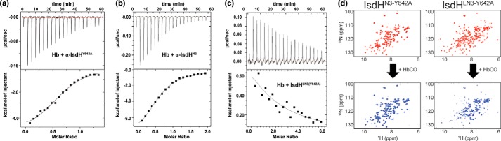
Staphylococcus aureus is a leading cause of life-threatening infections in the United States. It actively acquires the essential nutrient iron from human hemoglobin (Hb) using the iron-regulated surface-determinant (Isd) system. This process is initiated when the closely related bacterial IsdB and IsdH receptors bind to Hb and extract its hemin through a conserved tri-domain unit that contains two NEAr iron Transporter (NEAT) domains that are connected by a helical linker domain. Previously, we demonstrated that the tri-domain unit within IsdH (IsdHN2N3) triggers hemin release by distorting Hb's F-helix. Here, we report that IsdHN2N3 promotes hemin release from both the α- and β-subunits. Using a receptor mutant that only binds to the α-subunit of Hb and a stopped-flow transfer assay, we determined the energetics and micro-rate constants of hemin extraction from tetrameric Hb. We found that at 37 °C, the receptor accelerates hemin release from Hb up to 13,400-fold, with an activation enthalpy of 19.5 ± 1.1 kcal/mol. We propose that hemin removal requires the rate-limiting hydrolytic cleavage of the axial HisF8 Nϵ-Fe3+ bond, which, based on molecular dynamics simulations, may be facilitated by receptor-induced bond hydration. Isothermal titration calorimetry experiments revealed that two distinct IsdHN2N3·Hb protein·protein interfaces promote hemin release. A high-affinity receptor·Hb(A-helix) interface contributed ∼95% of the total binding standard free energy, enabling much weaker receptor interactions with Hb's F-helix that distort its hemin pocket and cause unfavorable changes in the binding enthalpy. We present a model indicating that receptor-introduced structural distortions and increased solvation underlie the IsdH-mediated hemin extraction mechanism.

Keywords: IsdB; IsdH; NEAT domain; bacterial pathogenesis; hemoglobin; iron-regulated surface determinant system; isothermal titration calorimetry (ITC); molecular dynamics; receptor; stopped-flow spectrophotometry.


Conflict of interest statement

The authors declare that they have no conflicts of interest with the contents of this article

Figures

Figure 1.
Figure 1.
S. aureus uses conserved tridomain-containing receptors to capture Hb. a, schematic showing the domain organization within the S. aureus Hb receptors, IsdH and IsdB. NEAT domains (N) that bind Hb and hemin (oxidized form of heme) are shown in gray and black, respectively. The helical domain that connects them is labeled linker. Residue numbers that define the boundaries of the functionally homologous NEAT domains are indicated. b, crystal structure of the Hb·α-IsdHN2N3 complex (PDB code 4XS0) with only contacts to the α-subunit shown. Proteins are shown in ribbon format, and the hemin group is represented by a space-filling model. Density for residues in the segment connecting the N2 and linker domains are absent in the electron density (dashed line). c, close-up view of the LN3·Hb interface that is distorted. α-IsdH is shown in blue, and ribbon diagram of αHb is shown in yellow (α-IsdH, residues Ala-326–Asp-660 from IsdH containing 365FYHYA369365YYHYF369 mutations). The F-helix in the receptor·Hb complex that is distorted (red) is overlaid with the native F-helix observed in the isolated Hb protein (green) (PDB code 2DN2). The hemin group is represented by a space-filling model (gray) with the iron atom in red.
Figure 2.
Figure 2.
Stoichiometry of Hb·receptor complexes determined by analytical ultracentrifugation. a, representative absorbance scans at 412 nm at equilibrium are plotted versus the distance from the axis of rotation for a mixture of Hb0.1 + IsdHY642A (blue circles) and Hb0.1 + α-IsdHY642A (red triangles). Hb0.1 was maintained in the carbonmonoxy-ligated state, and both receptors contained the Y642A mutation to prevent heme transfer. Protein samples were mixed at a ratio of 5 μm Hb0.1 and 150 μm receptor and were centrifuged at 25 °C for at least 48 h at 13,000 rpm. The solid lines represent the global nonlinear least-squares best fit of all the data to a single molecular species with a baseline fit. Residuals of the fit are shown above each panel. b, representative scans identical to the conditions described in a with the exception that 5 μm β-CO (isolated β-chains) was used, and samples were centrifuged at a speed of 9000 rpm.
Figure 3.
Figure 3.
Hemin transfer by various Hb and receptors species. a, spectral changes in the UV-visible spectrum of the reaction containing 5 μm Hb0.1 with buffer (solid black line), 150 μm IsdHN2N3 (dotted blue line), and 150 μm α-IsdH (solid red line). Spectral traces were recorded at 1000 s post-mixing. b, representative stopped-flow time courses showing absorbance changes at 405 nm (ΔA405) after mixing 5 μm Hb or Hb0.1 with 150 μm apo-receptor. The data were fit to a double-exponential equation to obtain kfast and kslow hemin transfer rates. Spectral traces were normalized for comparison using the following equation: y = (yty0)/(yt + y0). c and d show the measured kfast and kslow rate constants, respectively. Values were derived from the time courses in b. A two-way analysis of variance was used to access the significance in the difference of rates. NS means not significant (p values >0.05), and *** corresponds to p values <0.001.
Figure 4.
Figure 4.
Hemin transfer from isolated β-globin chains. Representative stopped-flow time courses show absorbance changes at 408 nm (ΔA408) after mixing 5 μm isolated Metβ-globin chains with 150 μm apo-receptor or 50 μm apo-Mb. Relative spectral traces are shown for comparison in which all starting absorbances were normalized to a value of 1. Only the first 10 s are shown as absorbance artifacts were observed after this time as a result of the slow process of apo-globin denaturation.
Figure 5.
Figure 5.
Hemin transfer to myoglobin and as function of receptor concentration. a, spontaneous hemin release from the receptor and Hb was measured using H64Y/V68F apomyoglobin. Time courses of the absorbance change at 600 nm (ΔA600) of a mixture of H64Y/V68F apomyoglobin (Mb) with Hb (black line), Hb0.1 (dark gray line), and IsdHN2N3 (light gray line) at a final concentration of 50 μm apo-Mb to 5 μm holo-protein. The receptor has higher affinity for hemin as compared with Hb or Hb0.1. b, plots of the observed rate constants, kfast (dark gray circles) and kslow (light gray circles), versus the ratio of [α-IsdH] to [Hb0.1]. The concentration of Hb0.1 was held constant at 5 μm. The observed values were obtained in experiments similar to those described in the legend in Fig. 3. For kfast, fits to the data using Equation 1 are shown.
Figure 6.
Figure 6.
Temperature dependence of hemin transfer. a, representative normalized stopped-flow time courses as function of ΔA405. They were obtained by mixing 5 μm Hb0.1 with 200 μm α-IsdH. Spectral traces were normalized using the following equation: y = (yty0)/(yt + y0). b, plot of ln(kfast/T) versus 1/T is shown. The observed kfast values were obtained in experiments similar to those described in the legend in Fig. 3. The correlation factor, R2, is indicated on the plot.
Figure 7.
Figure 7.
IsdH used two distinct interfaces to promote hemin transfer. a, representative ITC data for the titration of 200 μm α-IsdHY642A into 30 μm HbCO (heme/globin-chain basis). Injections were made at 180-s intervals at 25 °C. b, representative ITC data for the titration of 300 μm α-IsdHN2 into 30 μm HbCO. c, representative ITC data for the titration of 850 μm α-IsdHLN3-Y642A into 30 μm HbCO. The top panels show the time course of the titration (black) and baseline (red). The bottom panels show the integrated isotherms. ORIGIN software was used to fit the data to a single-site binding model to derive thermodynamic parameters. Receptors containing the N3 domain have a Y642A mutation that prevents hemin binding. d, NMR titration data showing the effects of Hb binding on 15N-labeled polypeptides that contain only the N3 domain (IsdHN3-Y642A, left) or the linker and N3 domains (IsdHLN3-Y642A, right). The receptors contain a Y642A mutation that prevents hemin binding. 1H-15N HSQC spectra of the proteins in the absence (top) or presence (bottom) of a 10-fold excess of HbCO tetramer are shown. Significant peak broadening is only observed when Hb is added to IsdHLN3-Y642A, indicating that linker and N3 domains bind Hb, whereas the N3 domain in isolation does not bind to Hb.
Figure 8.
Figure 8.
Results of molecular dynamics simulations. A, hemoglobin dimer (gold) was bound to hemin molecules (gray), and an IsdHN2N3 molecule (green and blue) was simulated, along with a system lacking IsdHN2N3 (not shown). B, radial distribution functions of solvent hydrogen (solid lines) and oxygen (dotted lines) atoms show that in the receptor-bound system, solvent molecules are more frequently found at closer locations to the Nϵ–Fe bond then in the receptor-free system. Analysis of simulations by a grid inhomogeneous solvation theory analysis showed that in IsdHN2N3-free systems (C and D) the number of hydration sites (cyan) surrounding the hemin group is limited; however, in the IsdHN2N3-bound systems (E and F) significantly more hydration sites are identified around the Nϵ–Fe bond.
Figure 9.
Figure 9.
Model of the hemin extraction mechanism. Shown is a proposed hemin transfer reaction coordinate diagram. The IsdHN2N3 receptor recognizes Hb via two energetically distinct interfaces (N2·Hb and LN3·Hb interfaces). Top, schematic showing the steps in hemin transfer. Bottom, relative free energies of the intermediates. Free energies calculated from this study are labeled along reaction coordinate diagram. Breakage of the axial Fe–Nϵ bond is presumably rate-limiting (step 3). The following color scheme was used: N2, green; linker-N3, blue; Hb α-subunit, gold.

References

    1. Klevens R. M., Morrison M. A., Nadle J., Petit S., Gershman K., Ray S., Harrison L. H., Lynfield R., Dumyati G., Townes J. M., Craig A. S., Zell E. R., Fosheim G. E., McDougal L. K., Carey R. B., et al. (2007) Invasive methicillin-resistant Staphylococcus aureus infections in the United States. JAMA 298, 1763–1771 10.1001/jama.298.15.1763 - DOI - PubMed
    1. Centers for Disease Control and Prevention (2013) Active Bacterial Core Surveillance (ABCs) Report Emerging Infections Program Network Methicillin-Resistant Staphylococcus aureus, Atlanta, GA
    1. Dukic V. M., Lauderdale D. S., Wilder J., Daum R. S., and David M. Z. (2013) Epidemics of community-associated methicillin-resistant Staphylococcus aureus in the United States: a meta-analysis. PLoS ONE 8, e52722 10.1371/journal.pone.0052722 - DOI - PMC - PubMed
    1. Ong S. T., Ho J. Z., Ho B., and Ding J. L. (2006) Iron-withholding strategy in innate immunity. Immunobiology 211, 295–314 10.1016/j.imbio.2006.02.004 - DOI - PubMed
    1. Haley K. P., and Skaar E. P. (2012) A battle for iron: host sequestration and Staphylococcus aureus acquisition. Microbes Infect. 14, 217–227 10.1016/j.micinf.2011.11.001 - DOI - PMC - PubMed

Publication types

MeSH terms