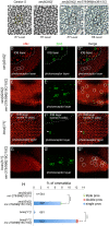
. 2018 Apr 9;145(7):dev159053.
doi: 10.1242/dev.159053.

The mir-279/996 cluster represses receptor tyrosine kinase signaling to determine cell fates in the Drosophila eye

Affiliations

The mir-279/996 cluster represses receptor tyrosine kinase signaling to determine cell fates in the Drosophila eye

Hong Duan et al. Development. .

Abstract

Photoreceptors in the crystalline Drosophila eye are recruited by receptor tyrosine kinase (RTK)/Ras signaling mediated by Epidermal growth factor receptor (EGFR) and the Sevenless (Sev) receptor. Analyses of an allelic deletion series of the mir-279/996 locus, along with a panel of modified genomic rescue transgenes, show that Drosophila eye patterning depends on both miRNAs. Transcriptional reporter and activity sensor transgenes reveal expression and function of miR-279/996 in non-neural cells of the developing eye. Moreover, mir-279/996 mutants exhibit substantial numbers of ectopic photoreceptors, particularly of R7, and cone cell loss. These miRNAs restrict RTK signaling in the eye, since mir-279/996 nulls are dominantly suppressed by positive components of the EGFR pathway and enhanced by heterozygosity for an EGFR repressor. miR-279/996 limit photoreceptor recruitment by targeting multiple positive RTK/Ras signaling components that promote photoreceptor/R7 specification. Strikingly, deletion of mir-279/996 sufficiently derepresses RTK/Ras signaling so as to rescue a population of R7 cells in R7-specific RTK null mutants boss and sev, which otherwise completely lack this cell fate. Altogether, we reveal a rare setting of developmental cell specification that involves substantial miRNA control.

Keywords: Drosophila; MicroRNA; R7 photoreceptor; RTK signaling.


Conflict of interest statement

Competing interestsThe authors declare no competing or financial interests.

Figures
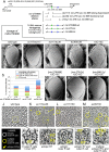
Fig. 1.
Fig. 1.
mir-279/996 alleles and corresponding adult Drosophila eye phenotypes. (A) The Drosophila mir-279/996 genomic region, along with three deletion alleles and three rescue transgenes built into a 16.6 kb genomic backbone. (B-I) Adult Drosophila eyes analyzed by scanning electron microscopy. (B) The wild-type eye exhibits a regular, crystalline organization. (C) A hypomorphic condition that is deleted for mir-279 and is impaired for miR-996 biogenesis appears externally normal, whereas genotypes that progressively remove miR-279/996 activity exhibit overt roughening and ommatidial disorganization (D-F). (G-I) The exterior eye phenotype caused by deletion of both mir-279 and mir-996 (in [15C] homozygotes) can be rescued by supplying both miRNAs (G), only mir-279 (H) or only mir-996 (I). (J-M) Plastic sections through adult eyes of wild type (J) and three heteroallelic mir-279/996 combinations (K-M). Normal ommatidia exhibit six large outer photoreceptor rhabdomeres (R1-6) surrounding a smaller inner photoreceptor rhabdomere (R7 or R8, depending on the apical-basal position). All of the transheterozygous mir-279/996 mutants exhibit populations of ommatidia with ectopic outer and/or inner photoreceptors, as annotated in the key to the left. Based on their position and morphology, the ectopic inner photoreceptors are predominantly R7. Some ommatidia can contain both ectopic outer and R7 photoreceptors, and the frequency of mutant ommatidia is noticeably higher in the strongest mutant transheterozygote ex36/15C (M). (N-R) Magnifications of individual ommatidia highlighting normal and different combinations of mutant photoreceptor identities. (S) Quantification of photoreceptor subtypes in the three heteroallelic mir-279/996 mutants examined. R7 is the predominant photoreceptor subtype affected in all genotypes.
Fig. 2.
Fig. 2.
Expression and activity of mir-279/996 in the developing pupal eye. (A) Transgenes to detect transcriptional activity or functional repression by mir-279/996; the former is a positive readout of miRNA expression, whereas the latter is a negative sensor of miRNA activity. (B) Schematic of ommatidial nuclei in the fly eye. Cone cell (CC) nuclei are located most apically, primary pigment cells and photoreceptors reside medially, and secondary and tertiary pigment cells and interommatidial bristle nuclei are located basally. (C-E″) Expression of the 16.6 kb mir-279/996-GFP transgene is readily detected throughout the cone cell layer as marked by DPax2 (C) and in the pigment cells and bristle cells (D), but is excluded from Elav+ photoreceptor neurons. (E) Magnification of the boxed region in D, highlighting exclusion of mir-279/996-GFP activity from Elav+ cells. (F-K‴) Functional repression detected by the tub-GFP-miR-279 sensor. Note that this is a cytoplasmic sensor, whereas the cell-specific markers are nuclear. (F) The tub-GFP-miR-279 sensor is largely excluded from the cone cell layer. (G) Magnified view of the boxed region in F. (H) The tub-GFP-miR-279 sensor is reactivated in cone cells when placed in the mir-279/996[ex36/ex36] background. (I) The tub-GFP-miR-279 sensor is coincident with Elav+ photoreceptors but is excluded from pigment cells. (J-J‴′′) Higher magnification view emphasizing the on and off spatial pattern of the miR-279 sensor in adjacent Elav+ and DPax2+ cells, respectively. (K-K‴) The tub-GFP-miR-279 sensor is reactivated in non-neuronal cells within the photoreceptor plane of mir-279/996[ex36/ex36] mutants.
Fig. 3.
Fig. 3.
Cell specification defects in mir-279/996 mutant eyes. Stainings and quantifications in A-S were from ∼45 h APF pupal eyes, while stainings in T-W were from adult eyes. (A-F) Arm labels the zonula adherens of apically constricted photoreceptors. (A) Seven of the eight neurons are labeled by Arm in a single optical section at the z-level in which the photoreceptor apices are well separated. (B) 15C homozygotes frequently have supernumerary photoreceptors, with ommatidia bearing eight Arm+ photoreceptors in a single optical section (green circles), and occasionally more (the yellow circle indicates an ommatidium with nine or possibly ten photoreceptors). (C) These defects were suppressed by a 16.6 kb mir-279/996 genomic transgene. (A′-C′) Higher magnifications of individual ommatidial groups (boxed regions in A-C) with photoreceptors labeled. A potential tenth photoreceptor in B′ is indicated with an asterisk. (D) In the cone cell layer, Arm labels groups of four cones in each wild-type ommatidium. (E) mir-279/996[15C/15C] mutants frequently have only three (or even two) cone cells (circles); very rarely, five cone cell clusters are seen (yellow circle). (F) Cone cell defects are fully rescued by the genomic transgene. (D′-F′) Higher magnifications of individual ommatidial groups (boxed regions in D-F) with cone cells labeled. (G,H) Quantification of ommatidia with aberrant photoreceptor number (G) or cone cell number (H) in mir-279/996 mutants and rescues. Error bars indicate s.d. ****P<0.0001, one-way ANOVA with Tukey's HSD post-hoc test. (I-K″) Co-staining for Dlg (green, cell membranes) and DPax2 (red, cone cell nuclei). (I) The regular pattern of four cone cells per ommatidium is seen in wild type (wt). (J) mir-279/996[15C/15C] mutants frequently exhibit three cone cells per ommatidium, which is rescued by a genomic transgene (K). (L-N) Staining for the neuronal marker Elav demonstrates mutant ommatidia with ectopic photoreceptors (asterisks). (O-Q) Co-staining for Elav and Pros shows that many ectopic photoreceptors are R7 cells. (R,R′) Region of mir-279/996 mutant eye that shows an especially high frequency of ectopic Pros photoreceptors (asterisks). (S) Quantification of ommatidia with ectopic Pros+ R7 cells. Error bars indicate s.d. ****P<0.0001, one-way ANOVA with Tukey’s HSD post-hoc test. (T-W) Staining for rhodopsins selectively expressed in terminally differentiated R7 cells: Rh3 (T,V) and Rh4 (U,W). mir-279/996 mutant ommatidia are labeled; white circles indicate two R7 cells and yellow circles indicate three R7 cells.
Fig. 4.
Fig. 4.
miR-279/996 directly repress multiple positive components of RTK/Ras pathways. (A) Among 3′ UTRs bearing target sites for the shared miR-279/996 seed sequence, three (ru, rho, boss) are positive components of RTK/Ras that promote photoreceptor/R7 specification. rho bears a 7mer-1A site, whereas boss and ru both contain high-affinity 8mer sites. (B) All of these sites are conserved across the 12 sequenced Drosophila genomes, as exemplified for ru. (C) Luciferase sensor assays demonstrate target 3′ UTR repression by miR-279 and miR-996. Hairless (H) is used as a negative control, while nerfin-1 is a positive control that is exceptionally well repressed by both miRNAs. rho, ru and boss are repressed 2- to 3-fold by ectopic miR-279/996 in a seed-dependent manner. Error bars indicate s.d. (D-G″) Evidence that miR-279 represses Ras pathway 3′ UTRs in vivo. Shown are the central portions of wing imaginal discs that express GFP ubiquitously (tub-GFP-3′ UTR sensors), in a genetic background in which miR-279 is ectopically expressed in a central stripe labeled by DsRed. (D) miR-279 does not affect a control GFP sensor, but induces cell-autonomous repression of GFP sensors linked to ru (E), rho (F) and boss (G) 3′ UTRs.
Fig. 5.
Fig. 5.
mir-279/996 mutant phenotypes are due to elevated Ras pathway activity. Except for the wild-type eye (A), all other adult and pupal eye samples are homozygous for mir-279/996[15C], with other heterozygous mutations as indicated. (A-G) Scanning electron microscopy of adult eyes. (A) Normal regular arrangement of wild-type ommatidia. The rough eye of the mir-279/996[15C] homozygote (B) is not modified by double heterozygosity for nerfin-1 and esg (C), but is partially suppressed by Egfr/+ (D) and ru/+ (E), and strongly suppressed by phyl/+ (F) and ru, rho/+, + (G). (H-S′) 45 h APF eyes stained for the indicated markers. (H-M) Arm staining focused on photoreceptor apices. Mutant ommatidia with ectopic photoreceptors (eight in one optical section) are circled; the inset (H) shows examples of ommatidia with seven and eight photoreceptors at higher magnification. The phenotype of [15C/15C] (H) is dominantly suppressed by heterozygosity for positive Ras pathway components (I-L) and dominantly enhanced by argos, a Ras pathway inhibitor (M). (N-S) Pros (green) and Elav (red) staining to detect R7 cells. The phenotype of [15C/15C] is not substantially modified by double heterozygosity for nerfin-1 and esg (N), but is dominantly suppressed by heterozygosity for positive Ras pathway components (O-R) and dominantly enhanced by argos (S). (T,U) Quantification of ectopic photoreceptor (T) and ectopic R7 (U) phenotypes in various genotypes. Error bars indicate s.d. **P<0.01, ***P<0.001, one-way ANOVA with Tukey's post-hoc test. (V) Retinal section of argos, 15C/15C adult eye illustrates a high frequency of ectopic R7 cells (arrowheads), which can be identified based on their central position within the circled ommatidia.
Fig. 6.
Fig. 6.
Deletion of mir-279/996 partially rescues R7 cells in sev and boss mutants. (A-C) Plastic sections of adult eyes. (A) Section of Canton S at the R7 cell level shows the characteristic trapezoidal arrangement of seven photoreceptor rhabdomeres in each ommatidia. (B) Section of sev[d2] mutant shows only six photoreceptors, with the centrally located R7 absent. (C) Example of rescued R7 cell in sev[d2]; mir-279/996[ex36/15C] eye; an adjacent section from the R8 level confirms the presence of R8 (C′), demonstrating that the cell assigned as R7 is not a misplaced R8. (D-G″) Double staining for Pros (green) and Elav (red) at 45 h APF. (D) sev mutant eye lacks Pros+ photoreceptors. Proper staining is confirmed by including a section that overlaps the interommatidial bristle (IOB) layer, where adjacent pairs of Pros+ sheath (S) and Elav+ neuronal (N) cells are present in each IOB organ. (E) sev; mir-279/996 double-mutant eye shows presence of Pros+ (R7) cells; their identity as photoreceptors is evidenced by colabeling for Elav (circled in E″). (F) boss mutant eye lacks Pros+ photoreceptors. (G) boss, mir-279/996 double-mutant eye exhibits rescue of a population of R7 cells (circled in G″). (H) Quantification of R7 rescue in sev and boss mutants by concomitant deletion of mir-279/996. Error bars indicate s.d. ****P<0.0001, one-way ANOVA with Tukey's post-hoc test.

Similar articles

Cited by

References

    1. Basler K., Christen B. and Hafen E. (1991). Ligand-independent activation of the sevenless receptor tyrosine kinase changes the fate of cells in the developing Drosophila eye. 64, 1069-1081. 10.1016/0092-8674(91)90262-W - DOI - PubMed
    1. Bejarano F., Bortolamiol-Becet D., Dai Q., Sun K., Saj A., Chou Y.-T., Raleigh D. R., Kim K., Ni J.-Q., Duan H. et al. (2012). A genome-wide transgenic resource for conditional expression of Drosophila microRNAs. 139, 2821-2831. 10.1242/dev.079939 - DOI - PMC - PubMed
    1. Brennecke J., Hipfner D. R., Stark A., Russell R. B. and Cohen S. M. (2003). bantam encodes a developmentally regulated microRNA that controls cell proliferation and regulates the proapoptotic gene hid in Drosophila. 113, 25-36. 10.1016/S0092-8674(03)00231-9 - DOI - PubMed
    1. Cagan R. L. and Ready D. F. (1989). Notch is required for successive cell decisions in the developing Drosophila retina. 3, 1099-1112. 10.1101/gad.3.8.1099 - DOI - PubMed
    1. Cayirlioglu P., Kadow I. G., Zhan X., Okamura K., Suh G. S., Gunning D., Lai E. C. and Zipursky S. L. (2008). Hybrid neurons in a microRNA mutant are putative evolutionary intermediates in insect CO2 sensory systems. 319, 1256-1260. 10.1126/science.1149483 - DOI - PMC - PubMed

Publication types

MeSH terms