. 2018 Jun;24(6):865-879.
doi: 10.1261/rna.063206.117. Epub 2018 Mar 14.

Transmission of microRNA antimiRs to mouse offspring via the maternal-placental-fetal unit

Affiliations

Transmission of microRNA antimiRs to mouse offspring via the maternal-placental-fetal unit

Jonas Hönig et al. RNA. 2018 Jun.

Abstract

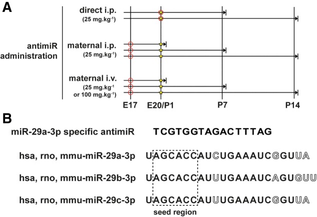
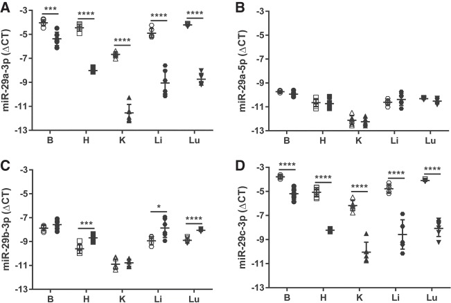
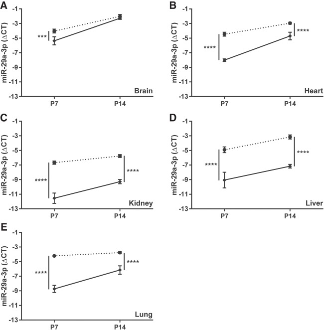
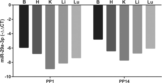
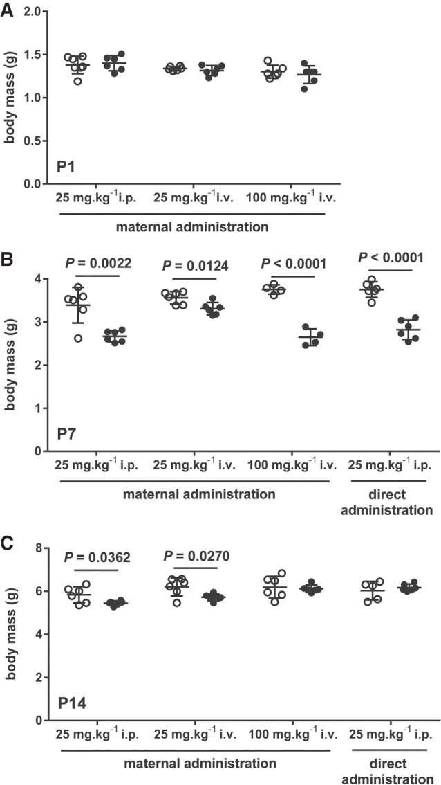
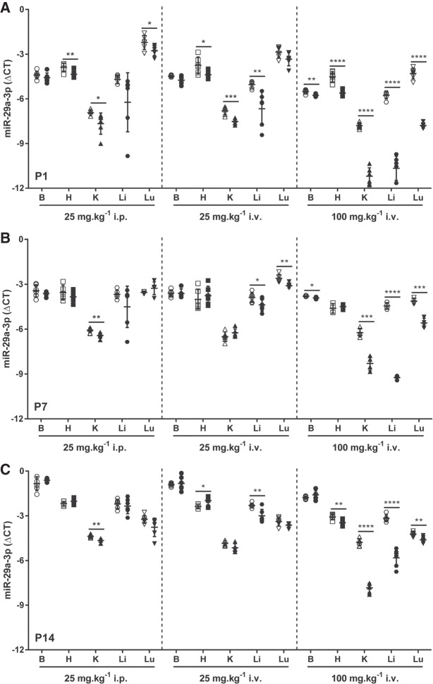
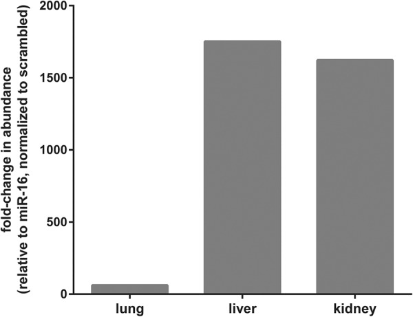
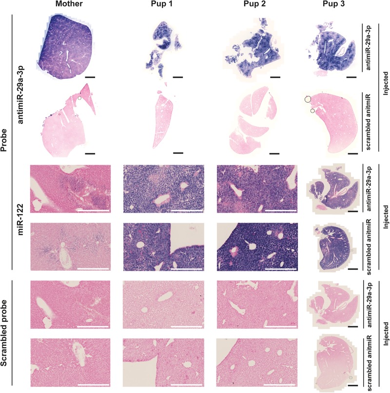
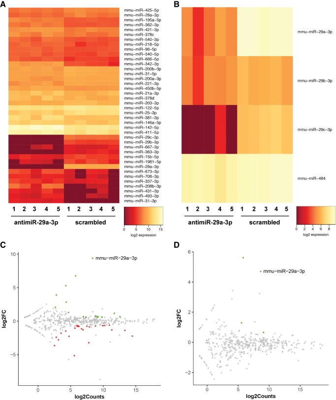
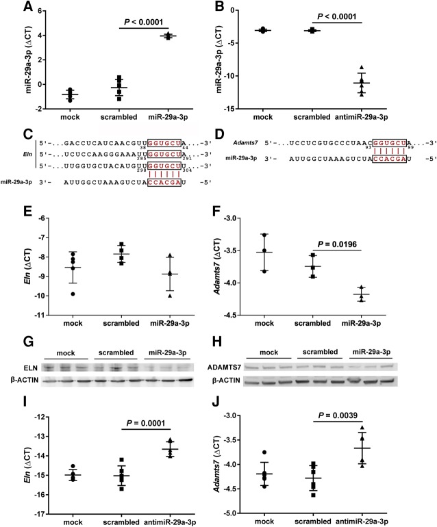
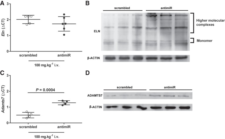
The emergence of microRNA as regulators of organogenesis and tissue differentiation has stimulated interest in the ablation of microRNA expression and function during discrete periods of development. To this end, inducible, conditional modulation of microRNA expression with doxycycline-based tetracycline-controlled transactivator and tamoxifen-based estrogen receptor systems has found widespread use. However, the induction agents and components of genome recombination systems negatively impact pregnancy, parturition, and postnatal development; thereby limiting the use of these technologies between late gestation and the early postnatal period. MicroRNA inhibitor (antimiR) administration also represents a means of neutralizing microRNA function in vitro and in vivo. To date, these studies have used direct (parenteral) administration of antimiRs to experimental animals. As an extension of this approach, an alternative means of regulating microRNA expression and function is described here: the maternal-placental-fetal transmission of antimiRs. When administered to pregnant dams, antimiRs were detected in offspring and resulted in a pronounced and persistent reduction in detectable steady-state free microRNA levels in the heart, kidney, liver, lungs, and brain. This effect was comparable to direct injection of newborn mouse pups with antimiRs, although maternal delivery resulted in fewer off-target effects. Furthermore, depletion of steady-state microRNA levels via the maternal route resulted in concomitant increases in steady-state levels of selected microRNA targets. This novel methodology permits the temporal regulation of microRNA function during late gestation and in neonates, without recourse to conventional approaches that rely on doxycycline and tamoxifen, which may confound studies on developmental processes.

Keywords: antimiR; development; maternal transmission; microRNA; organogenesis.


Figures

FIGURE 1.
FIGURE 1.
Schematic illustration of the antimiR administration protocol. (A) An antimiR directed against miR-29a-3p (or a scrambled antimiR) was administered by injection (○) via the intraperitoneal (i.p.) route (25 mg kg−1) on the day of birth [postnatal day (P)1; ⋆] to newborn pups; or via either the i.p. (25 mg kg−1) or intravenous (i.v.; 25 or 100 mg kg−1) routes to pregnant dams on the 17th day of gestation, here denoted embryonic day (E)17. Organs were harvested (→|) on P1, P7, or P14. (B) The antimiR-29a-3p sequence corresponded to part of the seed sequence (contained within the dashed-line box) of the miR-29 family of microRNA, and as such, targeted all three miR-29 family members in mice: miR-29a-3p, miR-29b-3p, and miR-29c-3p. Nucleotide base mismatches comparing the three miR-29a family members are indicated with hollow letters. (hsa) Homo sapiens; (mmu) Mus musculus; (rno) Rattus norvegicus.
FIGURE 2.
FIGURE 2.
The miR-29 family members can be targeted in mouse pups directly injected with an antimiR. Either a scrambled antimiR (open symbols) or an antimiR directed against miR-29a-3p (closed symbols) was administered by intraperitoneal injection (25 mg kg−1) to newborn mouse pups on the day of birth. Organs were harvested from mouse pups on the seventh day of life: (B) brain, (H) heart, (K) kidney, (Li) liver, (Lu) lung. Total RNA pools were screened by real-time RT-PCR for (A) miR-29a-3p, (B) miR-29a-5p, (C) miR-29b-3p, and (D) miR-29c-3p. The miR steady-state expression levels are described by the mean ΔCT ± SD. (n = 5–6 animals, per group; each symbol represents an individual animal), where ΔCT = CT(Rnu6) − CT(miR of interest). Mean values were compared between the scrambled versus miR-29a-3p-specific antimiR-treated mouse pups by unpaired Student's t-test. (*) P < 0.05; (***) P < 0.001; (****) P < 0.0001.
FIGURE 3.
FIGURE 3.
The impact of a directly injected antimiR directed against miR-29a-3p persists, but is slowly lost over the first fourteen days of postnatal life in mouse pups. Either a scrambled antimiR (closed circle, dotted line) or an antimiR directed against miR-29a-3p (closed diamond, solid line) was administered by intraperitoneal injection (25 mg kg−1) to newborn mouse pups on the day of birth. The (A) brain, (B) heart, (C) kidney, (D) liver, and (E) lung were harvested en bloc from mouse pups on postnatal day (P)7 or P14. The impact of antimiR-29a-3p administered at P1, on steady-state miR-29a-3p levels in these five organs was assessed at P7 and P14. The miR steady-state expression levels are described by the mean ΔCT ± SD. (n = 5–6 animals, per group; data points for each individual animal are omitted, for clarity), where ΔCT = CT(Rnu6) − CT(miR-29a-3p). Mean values were compared between the scrambled versus miR-29a-3p-specific antimiR-treated mouse pups by unpaired Student's t-test. (***) P < 0.001; (****) P < 0.0001.
FIGURE 4.
FIGURE 4.
Reduced steady-state free miR-29a-3p levels are detected in the organs of antimiR-29a-3p-injected dams. The steady-state free miR-29a-3p levels were assessed by real-time RT-PCR in the brain (B), heart (H), kidney (K), liver (Li), and lungs (Lu) of dams at postpartum day (PP)1 and PP14, after intravenous injection of either a scrambled antimiR (100 mg kg−1) or antimiR-29a-3p (100 mg kg−1) at gestational day 17. Data are presented as mean ΔΔCT, where ΔΔCT reflects ΔCT(antimiR-29a-3p treatment)–ΔCT(scrambled antimiR treatment), the negative value reflecting reduced detectable free miR-29a-3p in antimiR-29a-3p-injected dams. The original ΔCT values were obtained by ΔCT = CT(Rnu6) − CT(miR-29a-3p).
FIGURE 5.
FIGURE 5.
Maternal and direct administration of antimiR-29a-3p did impact postnatal growth of mouse pups. The body mass of mouse pups delivered by dams treated with intraperitoneal (i.p.) or intravenous (i.v.) scrambled antimiR (open circles) or antimiR-29a-3p (closed circles) was assessed on (A) the day of birth, postnatal day (P)1; as well as on (B) P7 and (C) P14. Additionally, the body mass of mouse pups directly injected at P1 (via the i.p. route) with scrambled antimiR (open circles) or antimiR-29a-3p (closed circles) was also assessed at (B) P7 and (C) P14. Data reflect mean body mass ± SD. (n = 5–6, per group). Mean values were compared between the scrambled versus miR-29a-specific antimiR-treated mouse pups by unpaired Student's t-test. All significant differences (P < 0.05) are illustrated, and the P-value is provided above the pair of data sets.
FIGURE 6.
FIGURE 6.
Steady-state levels of miR-29a-3p in mouse pups are influenced by administration of antimiR-29a-3p to pregnant dams. Either a scrambled antimiR (open symbols) or an antimiR directed against miR-29a-3p (closed symbols) was administered by intraperitoneal (25 mg kg−1) or intravenous (i.v.; 25 or 100 mg kg−1) injection to pregnant dams on the 17th day of gestation. Organs were harvested from mouse pups, either on the day of birth, postnatal day (P)1, or at P7 or P14: (B) brain, (H) heart, (K) kidney, (Li) liver, (Lu) lung. Total RNA pools were screened by real-time RT-PCR for steady-state miR-29a-3p levels in organs from pups at (A) P1, (B) P7, and (C) P14. The miR steady-state expression levels are described by the mean ΔCT ± SD. (n = 4–6 animals, per group; each symbol represents an individual animal), where ΔCT = CT(Rnu6) − CT(miR-29a-3p). Mean values were compared between the scrambled versus miR-29a-specific antimiR-treated mouse pups by unpaired Student's t-test. (*) P < 0.05; (**) P < 0.01; (***) P < 0.001; (****) P < 0.0001.
FIGURE 7.
FIGURE 7.
AntimiR-29a-3p was detected in selected organs of pups born to antimiR-29a-3p-treated dams by stem–loop real-time RT-PCR. Scrambled antimiR or antimiR-29a-3p was administered (100 mg kg−1, via the intravenous route) to pregnant dams on gestational day 17. The antimiR-29a-3p was detected by stem–loop real-time RT-PCR in total RNA pools from the lungs, liver, and kidneys of offspring at postnatal day 7. The data reflect the mean fold-change in the abundance of PCR product detected by stem–loop real-time RT-PCR, relative to miR-16, and normalized to the scrambled antimiR-treated group, as described in Materials and Methods.
FIGURE 8.
FIGURE 8.
AntimiR-29a-3p was detected in selected organs of pups born to antimiR-29a-3p-treated dams by in situ hybridization. AntimiR-29a-3p was administered (100 mg kg−1, via the intravenous route) to pregnant dams on gestational day 17. The antimiR-29a-3p was detected by in situ hybridization in the livers of offspring at postnatal day 7. Illustrated are liver sections from a dam (Mother) and from three different pups born to dams treated with either scrambled antimiR or with antimiR-29a-3p. AntimiR-29a-3p was detected with an in situ probe directed against antimiR-29a-3p, while a probe directed against the developmentally regulated miR-122 served as a positive control for the in situ hybridization reaction, and a scrambled in situ hybridization probe served as a negative control for the in situ hybridization reaction. A low-magnification view is illustrated for all samples from Pup 3, while higher-magnification views are illustrated for the dam (Mother), as well as Pup 1 and Pup 2. Black scale bars represent 2 mm, while white scale bars 400 µm.
FIGURE 9.
FIGURE 9.
Administration of antimiR-29a-3p had multiple off-target effects on free microRNA steady-state levels in directly injected pups, but limited off-target effects after maternal delivery. Either scrambled antimiR or antmiR-29a-3p was administered to pregnant mice at gestational day 17 (dose, 100 mg kg−1, intravenous) or directly to newborn mouse pups at postnatal day (P)1 (the day of birth; dose, 25 mg kg−1, intraperitoneal). Livers were harvested from offspring from injected pregnant mice and from directly injected pups, at P7. An RNA-seq screen was undertaken on microRNA pools from harvested livers. Complete RNA-seq data are available at the NCBI Gene Expression Omnibus under GEO Series accession number GSE110888. Heatmaps reflecting RNA-seq data are provided for (A) livers from directly injected pups, and (B) livers from offspring of injected pregnant dams. All microRNA species are illustrated that exhibited a false-discovery rate of <0.1, a minimum of five reads, and a fold-change of at least 1.5. In addition, M/A plots are provided for (C) livers from directly injected pups, and (D) livers from offspring of injected pregnant dams, to relate log2(fold-change) to log2(RNA-seq counts), where green spots indicate decreased transcript abundance in antimiR-29a-3p-treated groups, gray spots exhibit no change in transcript abundance comparing the antimiR-29a-3p-treated versus scrambled antimiR-treated groups, and red spots indicate increased transcript abundance in antimiR-29a-3p-treated groups.
FIGURE 10.
FIGURE 10.
Validation of Eln and Adamts7 mRNA as targets of miR-29a-3p in NIH/3T3 cells in vitro. The NIH/3T3 cells were mock-transfected, or were transfected with either a scrambled microRNA mimic, a miR-29a-3p mimic, or an antimiR-29a-3p for 48 h (all at 80 nM). Total RNA pools from NIH/3T3 cells were screened by real-time RT-PCR for steady-state levels of free miR-29a-3p, after (A) transfection of a miR-29a-3p mimic, or (B) an antimiR-29a-3p. In silico analyses revealed both the (C) Eln mRNA and (D) the Adamts7 mRNA to be candidate targets of miR-29a-3p; where the Eln mRNA 3′-untranslated region (UTR) contained three (8mer34–44, 8mer285–291, and 7mer-m8298–304) predicted miR-29a-3p binding-sites, and the Adamts7 mRNA 3′-UTR contained a single (7mer-A193–99) predicted miR-29a-3p binding-site. Therefore, total RNA pools from miR-29a-3p mimic-treated NIH/3T3 cells were also screened by real-time RT-PCR for steady-state expression levels of (E) Eln and (F) Adamts7. Total protein extracts from NIH/3T3 cells were screened by immunoblot for steady-state protein expression levels of (G) elastin (ELN) and (H) ADAM metallopeptidase with thrombospondin type 1 motif 7 (ADAMTS7), where β-ACTIN served as a loading control. Total RNA pools from antimiR-29a-3p-treated NIH/3T3 cells were also screened by real-time RT-PCR for steady-state expression levels of (I) Eln and (J) Adamts7. For RT-PCR studies, steady-state free miR or mRNA expression levels are described by the mean ΔCT ± SD. (n = 3–6 separate transfection experiments, per group; each symbol represents an independent transfection experiment, where some symbols may coalesce). For microRNA analyses, ΔCT = CT(Rnu6) – CT(miR-29a-3p), while for mRNA analyses, ΔCT = CT(Polr2a) − CT(gene of interest). Mean values were compared between treatment conditions by one-way ANOVA with Tukey's post hoc modification, and P-values are provided for scrambled versus miR/antimiR treatments.
FIGURE 11.
FIGURE 11.
Maternal transmission of antimiR-29a-3p to mouse pups increases steady-state expression levels of miR-29a-3p target-genes. Either a scrambled antimiR (open symbols) or an antimiR directed against miR-29a-3p (closed symbols) was administered by intravenous (i.v.) injection at a dose of 100 mg kg−1 to pregnant dams on the 17th day of gestation. The liver was harvested from mouse pups on postnatal day (P)7. Steady-state mRNA expression levels were assessed by real-time RT-PCR for the miR-29a-3p targets (A) Eln, and (C) Adamts7, described by the mean ΔCT ± SD. (n = 4–6 animals per group; each symbol represents an animal, where some symbols may coalesce), where ΔCT = CT(Polr2a) − CT(gene of interest). Mean values were compared between treatment conditions by unpaired Student's t-test. P-values are provided when P < 0.05. Total liver protein extracts from pups at P7 were screened by immunoblot for steady-state protein expression levels of (B) elastin (ELN) and (D) ADAM metallopeptidase with thrombospondin type 1 motif 7 (ADAMTS7), where β-ACTIN served as a loading control. For immunoblots, protein extracts from three scrambled antimiR-treated mice and three antimiR-29a-3p-treated mice were run side-by-side, on the same gel.

Similar articles

Cited by

References

    1. Agarwal V, Bell GW, Nam JW, Bartel DP. 2015. Predicting effective microRNA target sites in mammalian mRNAs. Elife 4: e05005. - PMC - PubMed
    1. Alejandre-Alcázar MA, Kwapiszewska G, Reiss I, Amarie OV, Marsh LM, Sevilla-Pérez J, Wygrecka M, Eul B, Köbrich S, Hesse M, et al. 2007. Hyperoxia modulates TGF-β/BMP signaling in a mouse model of bronchopulmonary dysplasia. Am J Physiol Lung Cell Mol Physiol 292: L537–L549. - PubMed
    1. Baek D, Villen J, Shin C, Camargo FD, Gygi SP, Bartel DP. 2008. The impact of microRNAs on protein output. Nature 455: 64–71. - PMC - PubMed
    1. Benjamini Y, Hochberg Y. 1995. Controlling the false discovery rate: a practical and powerful approach to multiple testing. J R Statist Soc B 57: 289–300.
    1. Braems G, Denys H, De Wever O, Cocquyt V, Van den Broecke R. 2011. Use of tamoxifen before and during pregnancy. Oncologist 16: 1547–1551. - PMC - PubMed

Publication types