. 2018 Apr 20;38(2):BSR20171717.
doi: 10.1042/BSR20171717. Print 2018 Apr 26.

Target identification reveals protein arginine methyltransferase 1 is a potential target of phenyl vinyl sulfone and its derivatives

Affiliations

Target identification reveals protein arginine methyltransferase 1 is a potential target of phenyl vinyl sulfone and its derivatives

Cheng-Han Yu et al. Biosci Rep. .

Abstract

Phenyl vinyl sulfone (PVS) and phenyl vinyl sulfonate (PVSN) inactivate protein tyrosine phosphatases (PTPs) by mimicking the phosphotyrosine structure and providing a Michael addition acceptor for the active-site cysteine residue of PTPs, thus forming covalent adducts between PVS (or PVSN) and PTPs. We developed a specific antiserum against PVS. This antiserum can be used in general antibody-based assays such as immunoblotting, immunofluorescence staining, and immunoprecipitation. Target identification through immunoprecipitation and mass spectrometry analysis reveals potential targets of PVS, mostly proteins with reactive cysteine residues or low-pKa cysteine residues that are prone to reversible redox modifications. Target identification of PVSN has been conducted because the anti-PVS antiserum can also recognize PVSN. Among the targets, protein arginine methyltransferase 1 (PRMT1), inosine-5'-monophosphate dehydrogenase 1, vimentin, and glutathione reductase (GR) were further confirmed by immunoprecipitation followed by immunoblotting. In addition, PVSN and Bay11-7082 inhibited GR activity, and PVS, PVSN, and Bay 11-7082 inhibited PRMT1 activity in in vitro assays. In addition, treatment of PVSN, Bay11-7082, or Bay 11-7085 in cultured HeLa cells can cause the quick decline in the levels of protein asymmetric dimethylarginine. These results indicate that the similar moiety among PVS, PVSN, Bay 11-7082, and Bay 11-7085 can be the key structure of lead compounds of PRMT1. Therefore, we expect to use this approach in the identification of potential targets of other covalent drugs.

Keywords: Bay11-7082; covalent drug; phenyl vinyl sulfonate; phenyl vinyl sulfone; protein arginine methyltransferase 1; target identification.


Conflict of interest statement

The authors declare that there are no competing interests associated with the manuscript.

Figures

Figure 1
Figure 1. Structures of phenyl vinyl sulfone (PVS) and its derivatives
Figure 2
Figure 2. Phenyl vinyl sulfone (PVS) as a covalent protein tyrosine phosphatase (PTP) inhibitor
HeLa cells were treated with various concentrations of PVS in PBS for 1 h, washed with PBS for three times, and lysed with RIPA lysis buffer containing 10 mM 1,4-dithioerythritol. The proteins in the lysate were examined by Coomassie blue G-250 staining (left panel), immunoblotting with anti-phosphotyrosine (middle panel), and anti-PVS (right panel) following SDS/PAGE and electrotransfer. The control groups were treated with the same volume of DMSO; α-PVS, anti-PVS; α-pY, anti-phosphotyrosine; CBB, Coomassie blue G-250.
Figure 3
Figure 3. Competition of PVS labeling by a PTP inhibitor pervanadate (PVD) in cellulo (A) and in vitro (B)
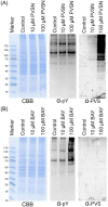
(A) HeLa cells were treated with 10 μM to 1 mM PVS in PBS for 1 h or with 0.1–2 mM PVD in PBS for 30 min. In the case of PVS and PVD combined treatment, HeLa cells were pretreated with PVD for 30 min and then treated with PVS in PBS for 1 h. The cells were then lysed with RIPA lysis buffer containing 10 mM 1,4-dithioerythritol. The proteins in the lysate were examined by Coomassie blue G-250 staining (left panel), immunoblotting with anti-phosphotyrosine (middle panel), and anti-PVS (right panel) following SDS/PAGE and electrotransfer. (B) HeLa cell lysates were treated with 10 μM to 1 mM PVS for 1 h. In the case of PVS and PVD combined treatment, HeLa cell lysates were pretreated with PVD for 30 min and then treated with PVS in PBS for 1 h. The reactions were then stopped by adding the same volume of RIPA buffer containing 10 mM 1,4-dithioerythritol. The proteins in the lysate were examined by Coomassie blue G-250 staining (left panel) anti-PVS (right panel) following SDS/PAGE and immunoblotting. The control groups were treated with the same volume of DMSO.
Figure 4
Figure 4. Applications of anti-PVS antiserum in immunofluorescence staining and immunoprecipitation experiments
(A) HeLa cells were cultured on coverslips for at least 24 h and treated with 0–100 μM PVS in PBS for 1 h. The cells were then processed for routine immunofluorescence staining with anti-PVS and DyLight 488 labeled anti-guinea pig IgG secondary antibodies. (B) HeLa cells were treated with 1 mM PVS in PBS for 1 h and then lysed with a lysis buffer suitable for immunoprecipitation and then processed for routine immunoprecipitation experiments with anti-PVS or with anti-PTP 1B. The immunoprecipitates were then examined by SDS/PAGE and immunoblotting with anti-PTP1B or with anti-PVS. The control groups were treated with the same volume of DMSO.
Figure 5
Figure 5. Use of anti-PVS in recognition of PVSN and Bay 11-7082 adducts
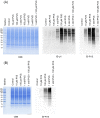
HeLa cells were treated with various concentrations of PVSN (A) or Bay 11-7082 (B) in PBS for 5 min, washed with PBS for three times, and lysed with RIPA lysis buffer containing 10 mM 1,4-dithioerythritol. The proteins in the lysate were examined by Coomassie blue G-250 staining, immunoblotting with anti-phosphotyrosine, and anti-PVS following SDS/PAGE and electrotransfer. The control groups were treated with the same volume of DMSO; α-PVS, anti-PVS; α-pY, anti-phosphotyrosine; CBB, Coomassie blue G-250.
Figure 6
Figure 6. Confirmation of PVS and PVSN tagging by immunoprecipitation followed by immunoblotting
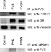
HeLa cells were treated with 500 μM PVS or PVSN in PBS for 5 min and the lysate was subjected to anti-PVS immunoprecipitation. The immunoprecipitate was then probed with anti-protein arginine methyltransferase 1 (PRMT1), anti-glutathione reductase (GR), and anti-vimentin (upper panel). For inosine-5′-monophosphate dehydrogenase 1 (IMPDH1), the lysate was immunoprecipitated with anti-IMPDH1 and then the immunoprecipitate was probed with anti-PVS (lower panel). The control groups were treated with the same volume of DMSO.
Figure 7
Figure 7. Effects of PVS, PVSN, and Bay 11-7082 on the in vitro enzyme activity of glutathione reductase
Recombinant glutathione reductase was first treated with NADPH and 10 or 50 μM PVS, PVSN, or Bay 11-7082 for 1 h at room temperature. In the control group, recombinant glutathione reductase was treated with NADPH and the same volume of DMSO used in the treatment of drugs in the assay buffer. Following the addition of oxidized glutathione, the decrease in absorbance at A340 was monitored over 10 min. Results were presented as mean of three independent experiments plus and minus standard deviation (compared with DMSO, *P<0.001.)
Figure 8
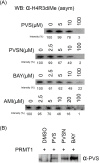
Figure 8. Effects of PVS, PVSN, Bay 11-7082, and AMI-1 on the in vitro enzyme activity of PRMT1 (A) and anti-PVS detection of PVS, PVSN, and Bay 11-7082 tagging of PRMT1 (B)
The reaction mixture contained 100 ng of recombinant PRMT1, 1 µg of full-length recombinant Histone H4, 1 µM S-adenosylmethionine, various concentrations of PVS, PVSN, Bay 11-7082, or AMI-1 (AMI) in a total volume of 100 µl in PBS, pH 7.4. DMSO was used in the control. The reaction was incubated at 37°C for 30 min and 10 µl of the reaction product was examined by SDS/PAGE and immunoblotting with anti-H4R3me2a for the recognition of Histone H4 asymmetric dimethylation at Arg3 (A) and anti-PVS (B). The 0 µM groups were treated with the same volume of DMSO.
Figure 9
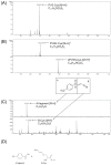
Figure 9. LC–MS analysis of reaction products of PVS, PVSN, and Bay 11-7082 with cysteine
In 1 ml of solution containing 100 μM cysteine, 100 μM PVS, PVSN, or Bay 11-7082, 20 mM NaHCO3 pH 8.4, the reaction was held at 37°C for 1 h. The reaction products were then analyzed by LC–ESI–MS. (A) The calculated molecular mass of PVS-cysteine adduct is 289 Da, and the calculated m/z [M + H]+ is 290 Da/e. The extracted ions of m/z 290.0528 Da/e represented PVS-cysteine adduct. (B) The calculated molecular mass of PVSN-cysteine adduct is 305 Da, and the calculated m/z [M+H]+ is 306 Da/e. The extracted ions of m/z 306.0479 Da/e represented PVSN-cysteine adduct. Extracted ions of m/z 490.0669 Da/e represented 2PVSN-cysteine because the calculated molecular mass of 2PVSN-cysteine adduct is 489 Da, and the calculated m/z [M + H]+ is 490 Da/e. (C) The extracted ions of m/z 155.0170 and 171.0230 Da/e represented Bay 11-7082 A fragment and B fragment-cysteine adduct respectively. The calculated molecular mass of A fragment-cysteine adduct is 156 Da (172 Da for B fragment-cysteine adduct), and the calculated m/z [M − H] is 155 Da/e (171 Da/e for B fragment-cysteine adduct). (D) The structures of Bay 11-7082 A fragment and B fragment-cysteine adduct were shown. Ions originating from original molecules by addition of a proton [M + H]+ or abstraction of a proton [M − H] were observed in positive or negative ion mode respectively.
Figure 10
Figure 10. Effects of PVS, PVSN, Bay 11-7082, Bay 11-7085, and AMI-1 on the signals of asymmetric dimethylarginine in HeLa Cells
HeLa cells were treated with 25 or 50 μM compounds in DMEM without FBS for 1 h. After washed with TBS for three times, the cells were then lysed and examined by SDS/PAGE and immunoblotting with anti-H4R3me2a antibody (α-H4R3diMe, asym) and with anti-dimethylarginine antibody (asymmetric) (α-diMeR, asym). The control groups were treated with the same volume of DMSO.

Similar articles

Cited by

References

    1. Tonks N.K. (2006) Protein tyrosine phosphatases: from genes, to function, to disease. Nat. Rev. Mol. Cell Biol. 7, 833–846 10.1038/nrm2039 - DOI - PubMed
    1. Barford D., Flint A.J. and Tonks N.K. (1994) Crystal structure of human protein tyrosine phosphatase 1B. Science 263, 1397–1404 10.1126/science.8128219 - DOI - PubMed
    1. Zhang Z.Y., Wang Y. and Dixon J.E. (1994) Dissecting the catalytic mechanism of protein-tyrosine phosphatases. Proc. Natl. Acad. Sci. U.S.A. 91, 1624–1627 10.1073/pnas.91.5.1624 - DOI - PMC - PubMed
    1. Lee S.R., Kwon K.S., Kim S.R. and Rhee S.G. (1998) Reversible inactivation of protein-tyrosine phosphatase 1B in A431 cells stimulated with epidermal growth factor. J. Biol. Chem. 273, 15366–15372 10.1074/jbc.273.25.15366 - DOI - PubMed
    1. Meng T.C., Fukada T. and Tonks N.K. (2002) Reversible oxidation and inactivation of protein tyrosine phosphatases in vivo. Mol. Cell 9, 387–399 10.1016/S1097-2765(02)00445-8 - DOI - PubMed

Publication types

MeSH terms