. 2018 Apr 11;38(15):3689-3707.
doi: 10.1523/JNEUROSCI.1587-17.2018. Epub 2018 Mar 14.

Functional MRI Responses to Passive, Active, and Observed Touch in Somatosensory and Insular Cortices of the Macaque Monkey

Affiliations

Functional MRI Responses to Passive, Active, and Observed Touch in Somatosensory and Insular Cortices of the Macaque Monkey

Saloni Sharma et al. J Neurosci. .

Abstract

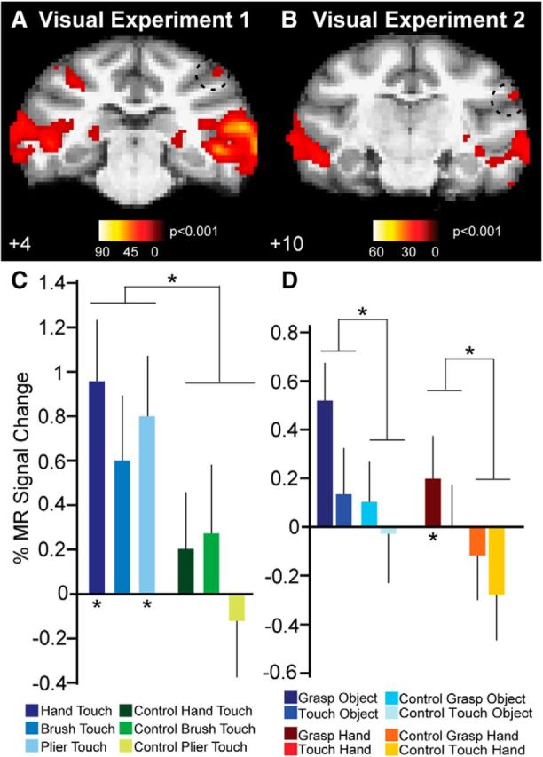
Neurophysiological data obtained in primates suggests that merely observing others' actions can modulate activity in the observer's motor cortices. In humans, it has been suggested that these multimodal vicarious responses extend well beyond the motor cortices, including somatosensory and insular brain regions, which seem to yield vicarious responses when witnessing others' actions, sensations, or emotions (Gazzola and Keysers, 2009). Despite the wealth of data with respect to shared action responses in the monkey motor system, whether the somatosensory and insular cortices also yield vicarious responses during observation of touch remains largely unknown. Using independent tactile and motor fMRI localizers, we first mapped the hand representations of two male monkeys' primary (SI) and secondary (SII) somatosensory cortices. In two subsequent visual experiments, we examined fMRI brain responses to (1) observing a conspecific's hand being touched or (2) observing a human hand grasping or mere touching an object or another human hand. Whereas functionally defined "tactile SI" and "tactile SII" showed little involvement in representing observed touch, vicarious responses for touch were found in parietal area PFG, consistent with recent observations in humans (Chan and Baker, 2015). Interestingly, a more anterior portion of SII, and posterior insular cortex, both of which responded when monkeys performed active grasping movements, also yielded visual responses during different instances of touch observation.SIGNIFICANCE STATEMENT Common coding of one's own and others' actions, sensations, and emotions seems to be widespread in the brain. Although it is currently unclear to what extent human somatosensory cortices yield vicarious responses when observing touch, even less is known about the presence of similar vicarious responses in monkey somatosensory cortex. We therefore localized monkey somatosensory hand representations using fMRI and investigated whether these regions yield vicarious responses while observing various instances of touch. Whereas "tactile SI and SII" did not elicit responses during touch observation, a more anterior portion of SII, in addition to area PFG and posterior insular cortex, all of which responded during monkeys' own grasping movements, yielded vicarious responses during observed touch.

Keywords: fMRI; grasping; macaque; motor; somatosensory; touch.


Figures

Figure 1.
Figure 1.
Functional MRI experiments and stimuli. A, To localize the hand representations in SI and SII responsive to passive cutaneous stimulation, we brushed the subject's left or right palm or side of the face, under light sedation in the scanner. Cutaneous stimulation blocks were interleaved with no stimulation (rest) blocks of equal duration (20 s). B, In an active touch localizer, subjects either grasped spheres of different diameters (Grasp) or placed their open right hands on a disk in front of them (Reach) in the dark inside the scanner. Motor blocks (30 s) were interleaved with baseline fixation blocks (Fix only), during which subjects were rewarded for fixating a red fixation target. C, In visual Experiment 1, we examined fMRI responses to observation of a conspecific's hand being touched by different effectors. Monkeys were required to fixate a small red fixation spot positioned on top of a monkey hand, while either a human hand (E), a brush (G), or a pair of closed pliers (I) touched the monkey's palm and swept down toward the fingertips. As controls, videos depicted the same effectors making a similar translation movement next to the monkey hand without touching it (F, H, J). D, In visual Experiment 2, we investigated fMRI responses to observing grasping and touching. Videos included an object being grasped (K) or touched with a finger (M), in addition to a human hand being grasped (O) or touched with a finger (Q). As controls, videos depicted the same hands making a similar translational movement next to the object or hand, without touching them (L, N, P, R).
Figure 2.
Figure 2.
Whole-brain fMRI responses during passive and active touch. A, B, Cutaneous stimulation of the monkeys' hands (compared with stimulation of the ipsilateral side of the face) yielded significant (p < 0.05, corr) fMRI responses in contralateral SI (tactile SI) and SII (tactile SII). Insets indicate fMRI responses for the same contrast at p < 0,001, uncorr. C, Active touch during grasp execution (compared with reach-only) in the dark yielded significant (p < 0.05, corr) contralateral responses in early visual, parietal, motor, premotor, prefrontal, somatosensory, and insular regions. White outlines indicate locations of tactile SI and SII regions responding during passive cutaneous stimulation in A and B. Insets, fMRI responses for the same contrast at p < 0,001, uncorr. D, In the ipsilateral hemisphere, grasping (compared with reach-only) yielded more restricted fMRI responses in similar early visual, parietal, motor, premotor, prefrontal, somatosensory, and insular regions.
Figure 3.
Figure 3.
Whole-brain fMRI responses during observation of touch. A, B, Observing a conspecific's hand being touched with different effectors (vs no-touch controls) yielded significant (p < 0.05, corr) responses in early visual, extrastriate, superior temporal sulcus (STS), parietal, premotor, prefrontal, somatosensory, and insular regions. Tactile SI and SII (white outlines) did not elicit significant responses to observation of a conspecific's hand being touched. Lowering the statistical threshold (p < 0.001, uncorr) confirmed the absence of touch-observation related responses (insets) in tactile SI and SII (white outlines). C, D, Observing an object or human hand being grasped or touched (vs no-touch controls) yielded significant (p < 0.05, corr) responses in similar early visual, extrastriate, STS, parietal, premotor, prefrontal, somatosensory, and insular regions. As for visual Experiment 1, tactile SI and tactile SII (white outlines) did not produce significant responses to observation of various instances of touch (vs controls). Lowering the statistical threshold (p < 0.001, uncorr) confirmed the absence of touch observation related responses (insets) in tactile SI and SII (white outlines).
Figure 4.
Figure 4.
Univariate fMRI responses to observed touch in tactile SI and SII regions-of-interest. A, B, Observing a conspecific's hand being touched by a human hand, brush or pair of pliers, in addition to no-touch control videos, did not yield significant visual responses (compared with fixation-only baseline) in tactile SI or SII in either hemisphere in any of the two monkey subjects (A, Monkey M1; B, Monkey M2). C, D, Observing a human hand grasping or merely touching either an object or another human hand, in addition to no-touch control videos, elicited no significant visual responses (compared with fixation-only baseline) in tactile SI or SII in either hemisphere in any of the two monkey subjects (C, Monkey M1; D, Monkey M2).
Figure 5.
Figure 5.
Multivariate fMRI decoding of observed touch in tactile SI and SII regions-of-interest. A, B, Classifier accuracies for binary decoding of observing a conspecific's hand being touched versus no-touch controls in tactile SI and SII of Monkey M1 (A) and Monkey M2 (B). Stippled lines indicate chance accuracy levels. Asterisk indicates significant decoding (p < 0.05, corr; see Materials and Methods). C, D, Classifier accuracies for binary decoding of observing a human hand grasping or touching an object or a hand (vs no-touch controls) in Monkey M1 (C) and Monkey M2 (D). Stippled lines indicate chance accuracy levels. All tested binary classifications yielded decoding accuracies not significantly different from chance level in tactile SI and SII, in both hemispheres of both monkeys.
Figure 6.
Figure 6.
Functional MRI responses to observed and active touch in upper bank of lateral sulcus of Monkey M1. A, B, Coronal section showing fMRI responses during observation of touch (vs no-touch controls) in visual Experiment 1 (A) and visual Experiment 2 (B) in upper bank of the lateral sulcus (stippled black outlines) in Monkey M1. C, Coronal section showing fMRI responses during active touch (grasping in the dark) in the same location in the upper bank of lateral sulcus in Monkey M1. D, E, Percentage MR signal changes (compared with fixation-only baseline) in Monkey M1 for observing a conspecific's hand being touched and no-touch control videos in left (D) and right (E) local maxima of visual activations of the upper bank of the lateral sulcus shown in A. Asterisks on top indicate a significant main effect for touch versus no touch (see Materials and Methods). Asterisks below horizontal axis indicate a significant response for each touch condition versus its respective no-touch control (Table 1). F, G, Percentage MR signal changes (compared with fixation-only baseline) in Monkey M1 for observing a human hand grasping or touching either an object or another hand and no-touch control videos in left (F) and right (G) local maxima of visual activations of the upper bank of the lateral sulcus shown in B. Asterisks on top indicate significant main effects for either touch versus no touch directed toward objects or directed toward hands. Asterisks below horizontal axis indicate significant response for each touch condition versus its respective no-touch control (Tables 2, 3).
Figure 7.
Figure 7.
Functional MRI responses to observed and active touch in upper bank of lateral sulcus of Monkey M2. A, B, Coronal section showing fMRI responses during observation of touch (vs no-touch controls) in visual Experiment 1 (A) and visual Experiment 2 (B) in upper bank of the lateral sulcus (stippled black outlines) in Monkey M2. C, Coronal section showing fMRI responses during active touch (grasping in the dark) in the same location in the upper bank of the lateral sulcus of Monkey M2. D, E, Percentage MR signal changes (compared with fixation-only baseline) in Monkey M2 for observing a conspecific's hand being touched and no-touch control videos in left (D) and right (E) local maxima of visual activations of the upper bank of the lateral sulcus shown in A. Asterisks on top indicate a significant main effect for touch versus no touch. Asterisks below horizontal axis indicate a significant response for each touch condition versus its respective no-touch control (Table 1). F, G, Percentage MR signal changes (compared with fixation-only baseline) in Monkey M2 for observing a human hand grasping or touching either an object or another hand and no-touch control videos, in left (F) and right (G) local maxima of visual activations of the upper bank of the lateral sulcus shown in B. Asterisks on top indicate significant main effects for either touch versus no touch directed toward objects or directed toward hands. Asterisks below horizontal axis indicate significant response for each touch condition versus its respective no-touch control (Tables 2, 3).
Figure 8.
Figure 8.
Functional MRI responses to observed and active touch in posterior insula of Monkey M1. A, B, Coronal section showing fMRI responses during observation of touch (vs no-touch controls) in visual Experiment 1 (A) and visual Experiment 2 (B) in posterior insula (stippled outlines) in Monkey M1. C, Coronal section showing fMRI responses during active touch (grasping in the dark) in in the same location in the posterior insula in Monkey M1. D, E, Percentage MR signal changes (compared with fixation-only baseline) in Monkey M1 for observing a conspecific's hand being touched and no-touch control videos in left (D) and right (E) local maxima of posterior insula activations shown in A. Asterisks on top indicate a significant main effect for touch versus no touch. Asterisks below horizontal axis indicate a significant response for each touch condition versus its respective no-touch control (Table 1). F, Percentage MR signal changes (compared with fixation-only baseline) in Monkey M1 for observing a human hand grasping or touching an object or another human hand, and no-touch control videos in right local maximum of posterior insula activation shown in B. Asterisks on top indicate significant main effects for either touch versus no touch directed toward objects or directed toward hands. Asterisks below horizontal axis indicate significant response for each touch condition versus its respective no-touch control (Tables 2, 3).
Figure 9.
Figure 9.
Functional MRI responses to observed and active touch in posterior insula of Monkey M2. A, B, Coronal section showing fMRI responses during observation of touch (vs no-touch controls) in visual Experiment 1 (A) and visual Experiment 2 (B) in posterior insula (stippled outlines) in Monkey M2. C, Coronal section showing fMRI responses during active touch (grasping in the dark) in the same location in the posterior insula of Monkey M2. D, E, Percentage MR signal changes (compared with fixation-only baseline) in Monkey M2 for observing a conspecific's hand being touched and no-touch control videos in left (D) and right (E) local maxima of posterior insula activations shown in A. Asterisks on top indicate a significant main effect for touch versus no touch. Asterisks below horizontal axis indicate a significant response for each touch condition versus its respective no-touch control (Table 1). F, G, Percentage MR signal changes (compared with fixation-only baseline) in Monkey M2 for observing a human hand grasping or touching an object or another human hand, and no-touch control videos in left (F) and right (G) local maxima of posterior insula activation shown in B. Asterisks on top indicate significant main effects for either touch versus no touch directed toward objects or directed toward hands. Asterisks below horizontal axis indicate significant response for each touch condition versus its respective no-touch control (Tables 2, 3).
Figure 10.
Figure 10.
Functional MRI responses to observed touch area PFG of Monkey M1. A, B, Coronal section showing fMRI responses during observation of touch (vs no-touch controls) in visual Experiment 1 (A) and visual Experiment 2 (B) in area PFG (stippled outlines) in Monkey M1. C, Percentage MR signal changes (compared with fixation-only baseline) in Monkey M1 for observing a conspecific's hand being touched and no-touch control videos in right local maximum of area PFG activation shown in A. Asterisks on top indicate a significant main effect for touch versus no touch. Asterisks below horizontal axis indicate a significant response for each touch condition versus its respective no-touch control (Table 1). D, Percentage MR signal changes (compared with fixation-only baseline) in Monkey M1 for observing a human hand grasping or touching an object or another human hand, and no-touch control videos in right local maximum of area PFG activation shown in B. Asterisks on top indicate significant main effects for either touch versus no touch directed toward objects or directed toward hands. Asterisks below horizontal axis indicate significant response for each touch condition versus its respective no-touch control (Tables 2, 3).
Figure 11.
Figure 11.
Functional MRI responses to observed and active touch in area PFG of Monkey M2. A, B, Coronal section showing fMRI responses during observation of touch (vs no-touch controls) in visual Experiment 1 (A) and visual Experiment 2 (B) in area PFG (stippled outlines) in Monkey M2. C, Percentage MR signal changes (compared with fixation-only baseline) in Monkey M2 for observing a conspecific's hand being touched and no-touch control videos in right local maximum of area PFG activation shown in A. Asterisks on top indicate a significant main effect for touch versus no touch. Asterisks below horizontal axis indicate a significant response for each touch condition versus its respective no-touch control (Table 1). D, E, Percentage MR signal changes (compared with fixation-only baseline) in Monkey M2 for observing a human hand grasping or touching an object or another human hand, and no-touch control videos in left (D) and right (E) local maxima of area PFG activations shown in B. Asterisks on top indicate significant main effects for either touch versus no touch directed toward objects or directed toward hands. Asterisks below horizontal axis indicate significant response for each touch condition versus its respective no-touch control (Tables 2, 3).
Figure 12.
Figure 12.
Functional MRI responses to passive, active and observed touch along the upper bank of the lateral sulcus. AD, Line plots showing MR percentage signal change related to passive touch (cutaneous stimulation of contralateral hand vs face; blue line), active touch (grasp vs reach-only execution with the right hand; green line), and observed touch (Experiment 2, object grasp observation vs no-touch control; red line), along the length of the upper bank of the lateral sulcus in left and right hemispheres of Monkeys M1 (A, B) and M2 (C, D). On the abscissa, the y-coordinates indicate the posterior–anterior extent of the path, starting near the posterior (p) side at the level of area PFG, toward the anterior (a) end of the lateral sulcus, as indicated in the sagittal insets. Shaded regions flanking the line plots indicate variability across runs.

Similar articles

Cited by

References

    1. Blakemore SJ, Bristow D, Bird G, Frith C, Ward J (2005) Somatosensory activations during the observation of touch and a case of vision-touch synaesthesia. Brain 128:1571–1583. 10.1093/brain/awh500 - DOI - PubMed
    1. Bonini L, Rozzi S, Serventi FU, Simone L, Ferrari PF, Fogassi L (2010) Ventral premotor and inferior parietal cortices make distinct contribution to action organization and intention understanding. Cereb Cortex 20:1372–1385. 10.1093/cercor/bhp200 - DOI - PubMed
    1. Borra E, Gerbella M, Rozzi S, Luppino G (2017) The macaque lateral grasping network: a neural substrate for generating purposeful hand actions. Neurosci Biobehav Rev 75:65–90. 10.1016/j.neubiorev.2017.01.017 - DOI - PubMed
    1. Bremmer F, Schlack A, Shah NJ, Zafiris O, Kubischik M, Hoffmann K, Zilles K, Fink GR (2001) Polymodal motion processing in posterior parietal and premotor cortex: a human fMRI study strongly implies equivalencies between humans and monkeys. Neuron 29:287–296. 10.1016/S0896-6273(01)00198-2 - DOI - PubMed
    1. Bufalari I, Aprile T, Avenanti A, Di Russo F, Aglioti SM (2007) Empathy for pain and touch in the human somatosensory cortex. Cereb Cortex 17:2553–2561. 10.1093/cercor/bhl161 - DOI - PubMed

Publication types

LinkOut - more resources