. 2018 Apr 15;200(8):2748-2756.
doi: 10.4049/jimmunol.1701492. Epub 2018 Mar 14.

Intracellular Nucleic Acid Sensing Triggers Necroptosis through Synergistic Type I IFN and TNF Signaling

Affiliations

Intracellular Nucleic Acid Sensing Triggers Necroptosis through Synergistic Type I IFN and TNF Signaling

Michelle Brault et al. J Immunol. .

Abstract

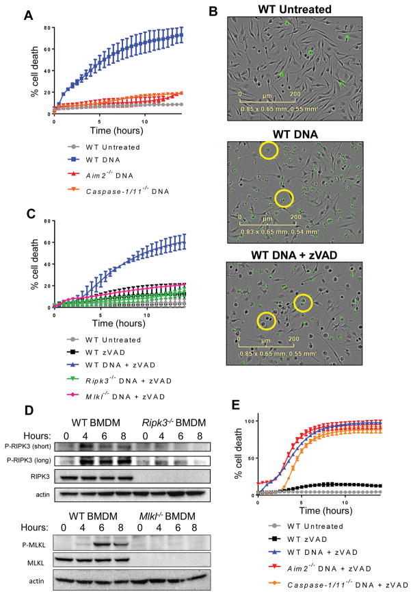
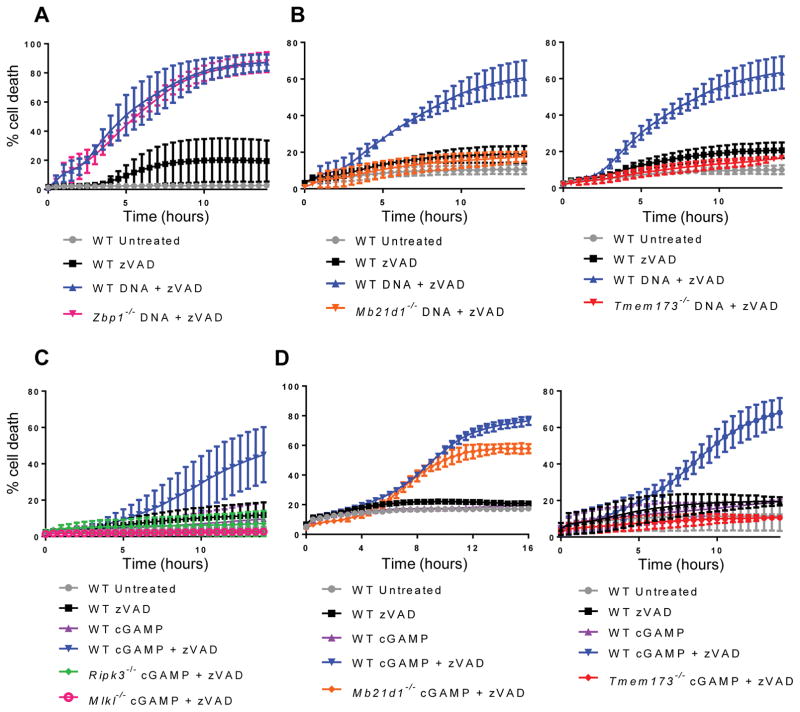
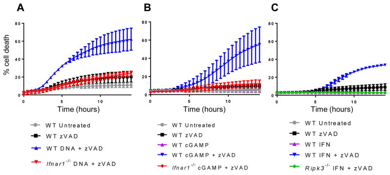
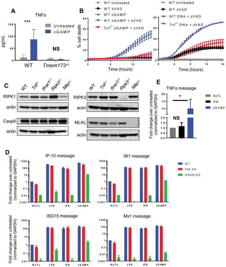
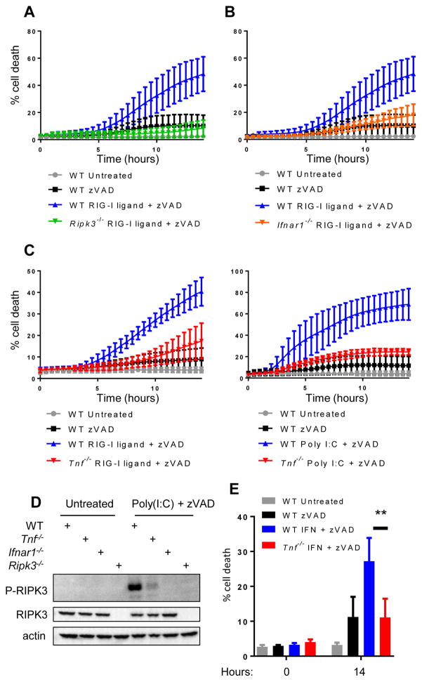
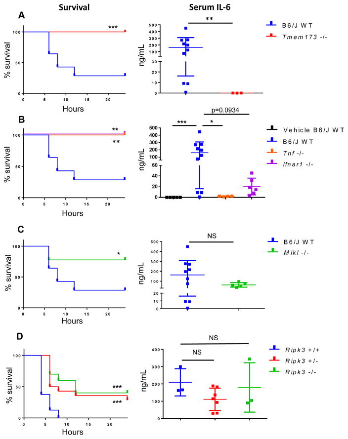
The sensing of viral nucleic acids within the cytosol is essential for the induction of innate immune responses following infection. However, this sensing occurs within cells that have already been infected. The death of infected cells can be beneficial to the host by eliminating the virus's replicative niche and facilitating the release of inflammatory mediators. In this study, we show that sensing of intracellular DNA or RNA by cGAS-STING or RIG-I-MAVS, respectively, leads to activation of RIPK3 and necroptosis in bone marrow-derived macrophages. Notably, this requires signaling through both type I IFN and TNF receptors, revealing synergy between these pathways to induce cell death. Furthermore, we show that hyperactivation of STING in mice leads to a shock-like phenotype, the mortality of which requires activation of the necroptotic pathway and IFN and TNF cosignaling, demonstrating that necroptosis is one outcome of STING signaling in vivo.


Conflict of interest statement

Disclosures

The authors declare no competing interests.

Figures

Figure 1
Figure 1. Introduction of DNA into the cytosol can trigger necroptotic cell death
(A) Kinetic cell death of WT, Aim2−/−, or Caspase-1/11−/− primary bone marrow-derived macrophages (BMDM) after transfection with 2 μg/ml DNA. Death measured by uptake of cell impermeable Sytox green dye, and normalized to starting number of cells stained with cell permeable Syto green dye to calculate percent cell death. Error bars represent SD from three independent experiments. (B) IncuCyte images of WT BMDM treated with 2 μg/ml DNA, or 2 μg/ml DNA + 50 μM zVAD. Sytox Green staining is shown in green. (C) Kinetic cell death of WT, Ripk3−/−, or Mlkl−/− BMDM after treatment with 2 μg/ml cytosolic DNA and 50 uM pan-caspase inhibitor zVAD. (D) Western blot analysis of phosphorylated RIPK3 or MLKL at short and long exposure following treatment of 5 μg/ml cytosolic DNA and 50 μM pan-caspase inhibitor zVAD in WT, Ripk3−/−, or Mlkl−/− BMDM. “z” in hours indicates zVAD alone control. (E) Kinetic cell death of WT, Aim2−/−, or Casp1/11−/− BMDM after treatment with 2 μg/ml cytosolic DNA and 50 μM pan-caspase inhibitor zVAD.
Figure 2
Figure 2. Cytosolic DNA triggers necroptosis via the cGAS-STING pathway
(A) Kinetic cell death of WT or Zbp1−/− BMDM after treatment with 2 μg/ml cytosolic DNA and 50 μM pan-caspase inhibitor zVAD. (B) Kinetic cell death of WT, Mb21d1−/−, Tmem173−/− BMDM after treatment with 2 μg/ml cytosolic DNA and 50 μM pan-caspase inhibitor zVAD. (C) Kinetic cell death of WT, Ripk3−/−, or Mlkl−/− BMDM after treatment with 2 μg/ml cGAMP and 25 μM pan-caspase inhibitor zVAD. (D) Kinetic cell death of WT, Mb21d1−/−, or Tmem173−/− BMDM after treatment with 2 μg/ml cGAMP and 25 μM pan-caspase inhibitor zVAD.
Figure 3
Figure 3. STING-dependent IFN production is necessary but not sufficient to induce necroptosis
(A) Kinetic cell death of WT or Ifnar1−/− BMDM after treatment with 2 μg/ml cytosolic DNA and 50 μM pan-caspase inhibitor zVAD. (B) Kinetic cell death of WT or Ifnar1−/− BMDM after treatment with 2 μg/ml cGAMP and 25 μM pan-caspase inhibitor zVAD. (C) Kinetic cell death of WT or Ripk3−/− BMDM after treatment with 100 units/ml recombinant IFN and 25 μM pan-caspase inhibitor zVAD.
Figure 4
Figure 4. STING-dependent TNF production is required for the induction of necroptosis
Luminex analysis of TNFα protein levels in supernatants from WT or Tmem173−/− BMDM following treatment with 5 μg/ml cGAMP. Error bars represent SD for three independent experiments. (B) Kinetic cell death of WT or Tnf−/− BMDM after treatment with 2 μg/ml cGAMP and 25 μM pan-caspase inhibitor zVAD, or 1 μg/ml cytosolic DNA and 50 μM pan-caspase inhibitor zVAD. (C) Western blot analysis of RIPK1, Caspase-8, RIPK3, and MLKL protein levels at steady state in WT, Tnf−/−, Ifnar1−/−, Ripk3−/−, or Mlkl−/− BMDM. (D) Quantitative PCR analysis of interferon stimulated genes (IP-10, Ifit1, ISG15, Mx1) in WT, Tnf−/−, or Ifnar1−/− BMDM following 6 hour treatment with 200 ng/ml LPS, 100 units/ml recombinant IFN, or 5 μg/ml cGAMP. Error bars represent SD for two independent experiments with three technical replicates. Raw values were normalized to housekeeping gene Gapdh and then to the WT untreated sample. (E) Quantitative PCR analysis of Tnf in WT BMDM following 6 hour treatment with 100 units/ml recombinant IFN or 5 μg/ml cGAMP. *p < 0.05, **p < 0.01, ***p < 0.001
Figure 5
Figure 5. Synergistic TNF and IFN singaling is required for necroptosis downstream of TLR3 and RIG-I
(A) Kinetic cell death of WT or Ripk3−/− BMDM after treatment with 1 μg/ml 5′-tri-phosphate RNA (RIG-I ligand) and 25 μM pan-caspase inhibitor zVAD. (B) Kinetic cell death of WT or Ifnar1−/− BMDM after treatment with 1 μg/ml tri-phosphate RNA (RIG-I ligand) and 25 μM pan-caspase inhibitor zVAD. (C) Kinetic cell death of WT or Tnf−/− BMDM after treatment with 1 μg/ml tri-phosphate RNA (RIG-I ligand) and 25 μM pan-caspase inhibitor zVAD or 1 μg/ml poly(I:C) and 25 μM pan-caspase inhibitor zVAD. (D) Western blot analysis of phosphorylated RIPK3 in WT, Tnf−/−, Ifnar1−/−, or Ripk3−/−BMDM following treatment with 2 μg/ml poly(I:C). Cells were harvested 8 hours post treatment. (E) Cell death analysis of WT or Tnf−/− BMDM after treatment with 100 units/ml recombinant IFN and 25 μM pan-caspase inhibitor zVAD.
Figure 6
Figure 6. STING agonists induce sterile shock that engages TNF, IFN, and necroptosis in vivo
(A) Survival analysis and serum IL-6 of age/sex matched WT C57BL/6J and Tmem173−/− mice after intraperitoneal injection of 40 mg/kg DMXAA. Serum was collected 6 hours post treatment. (B) Survival analysis and serum IL-6 of age/sex matched WT C57BL/6J, Tnf−/−, and Ifnar1−/− mice after intraperitoneal injection of 40 mg/kg DMXAA. (C) Survival analysis and serum IL-6 of age/sex matched WT C57BL/6J and Mlkl−/− mice after intraperitoneal injection of 40 mg/kg DMXAA. (D) Survival analysis and serum IL-6 of littermate C57BL/6N Ripk3+/+, Ripk3+/, Ripk3−/− mice after intraperitoneal injection of 40 mg/kg DMXAA. *p < 0.05, **p < 0.01, ***p < 0.001. All data is pooled from three or more independent experiments. B6/J WT data is pooled from all experiments and separated to different panels for ease of data comparison within panel.

Similar articles

Cited by

References

    1. Linkermann A, Green DR. Necroptosis. N Engl J Med. 2014;370:455–465. - PMC - PubMed
    1. Upton JW, Kaiser WJ, Mocarski ES. DAI/ZBP1/DLM-1 complexes with RIP3 to mediate virus-induced programmed necrosis that is targeted by murine cytomegalovirus vIRA. Cell Host and Microbe. 2012;11:290–297. - PMC - PubMed
    1. Upton JW, Kaiser WJ, Mocarski ES. Virus Inhibition of RIP3-Dependent Necrosis. Cell Host and Microbe. 2010;7:302–313. - PMC - PubMed
    1. Nogusa S, Thapa RJ, Dillon CP, Liedmann S, Oguin TH, Ingram JP, Rodriguez DA, Kosoff R, Sharma S, Sturm O, Verbist K, Gough PJ, Bertin J, Hartmann BM, Sealfon SC, Kaiser WJ, Mocarski ES, López CB, Thomas PG, Oberst A, Green DR, Balachandran S. RIPK3 Activates Parallel Pathways of MLKL-Driven Necroptosis and FADD-Mediated Apoptosis to Protect against Influenza A Virus. Cell Host and Microbe. 2016;20:13–24. - PMC - PubMed
    1. Huang Z, Wu SQ, Liang Y, Zhou X, Chen W, Li L, Wu J, Zhuang Q, Chen C, Li J, Zhong CQ, Xia W, Zhou R, Zheng C, Han J. RIP1/RIP3 binding to HSV-1 ICP6 initiates necroptosis to restrict virus propagation in mice. Cell Host and Microbe. 2015;17:229–242. - PubMed

Publication types

MeSH terms