. 2018 May 14;92(11):e01999-17.
doi: 10.1128/JVI.01999-17. Print 2018 Jun 1.

Positive Regulation of Hepatitis E Virus Replication by MicroRNA-122

Affiliations

Positive Regulation of Hepatitis E Virus Replication by MicroRNA-122

Bangari Haldipur et al. J Virol. .

Abstract

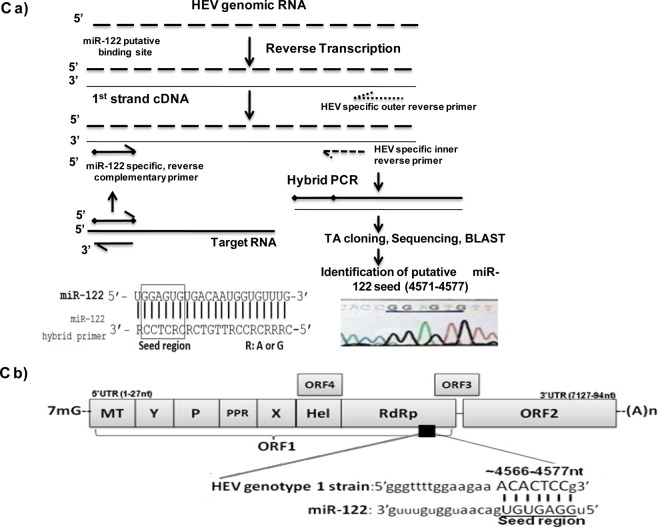
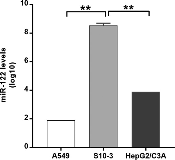
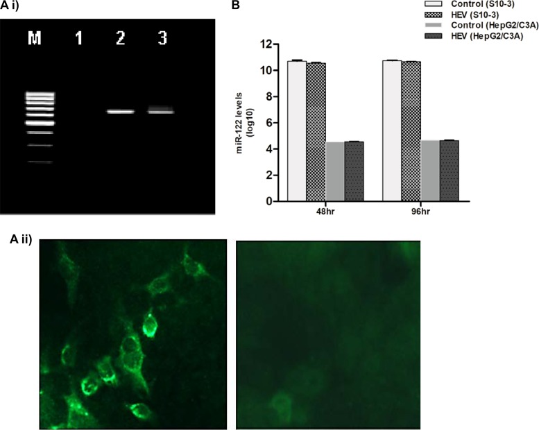
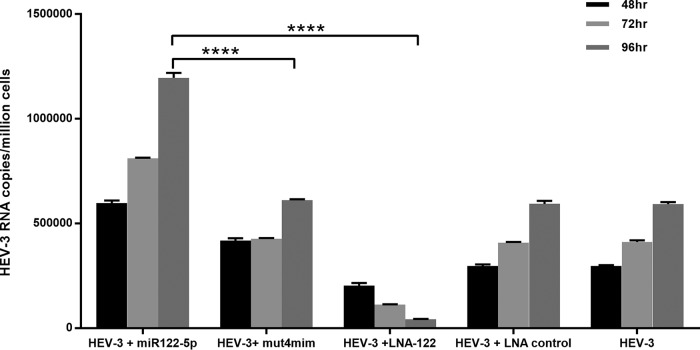
The molecular mechanisms of liver pathology and clinical disease in hepatitis E virus (HEV) infection remain unclear. MicroRNAs (miRNAs) are known to modulate viral pathogenesis either by directly altering viral gene expression or by enhancing cellular antiviral responses. Given the importance of microRNA-122 (miR-122) in liver pathobiology, we investigated possible role of miR-122 in HEV infection. In silico predictions using HEV genotype 1 (HEV-1), HEV-2, HEV-3, and HEV-4 sequences showed that the majority of genomes (203/222) harbor at least one miR-122/microRNA-122-3p (miR-122*) target site. Interestingly, HEV-1 genomes showed a highly (97%) conserved miR-122 target site in the RNA-dependent RNA polymerase (RdRp) region (RdRpc). We analyzed the significance of miR-122 target sites in HEV-1/HEV-3 (HEV-1/3) genomes by using a replicon-based cell culture system. HEV infection did not change the basal levels of miR-122 in hepatoma cells. However, transfection of these cells with miR-122 mimics enhanced HEV-1/3 replication and depletion of miR-122 with inhibitors led to suppression of HEV-1/3 replication. Mutant HEV-1 replicons with an altered target RdRpc sequence (CACTCC) showed a drastic decrease in virus replication, whereas introduction of alternative miR-122 target sites in mutant replicons rescued viral replication. There was enrichment of HEV-1 RNA and miR-122 molecules in RNA-induced silencing complexes in HEV-infected cells. Furthermore, pulldown of miR-122 molecules from HEV-infected cells resulted in pulldown of HEV genomic RNA along with miR-122 molecules. These observations indicate that miR-122 facilitates HEV-1 replication, probably via direct interaction with a target site in the viral genome. The positive role of miR-122 in viral replication presents novel opportunities for antiviral therapy and management of hepatitis E.IMPORTANCE Hepatitis E is a problem in both developing and developed countries. HEV infection in most patients follows a self-limited course; however, 20% to 30% mortality is seen in infected pregnant women. HEV superinfections in patients with chronic hepatitis B or hepatitis C virus infections are associated with adverse clinical outcomes, and both conditions warrant therapy. Chronic HEV infections in immunocompromised transplant recipients are known to rapidly progress into cirrhosis. Currently, off-label use of ribavirin (RBV) and polyethylene glycol-interferon (PEG-IFN) as antiviral therapy has shown promising results in both acute and chronic hepatitis E patients; however, the teratogenicity of RBV limits its use during pregnancy, while alpha IFN (IFN-α) increases the risk of transplant rejections. Experimental data determined with genotype 1 virus in the current study show that miR-122 facilitates HEV replication. These observations present novel opportunities for antiviral therapy and management of hepatitis E.

Keywords: Ago2; hepatitis E virus; interaction; miR-122; target site; virus replication.


Figures

FIG 1
FIG 1
(A) Computational prediction of miR-122/miR-122* targets in the HEV genomes. The HEV genomes were screened for putative miR-122/miR-122* target sites using RegRNA, and the prediction patterns were analyzed. The results of the analysis are depicted. (B) Conserved miR-122 target sites in the HEV-1 genome. The replicon developed from the highlighted sequence was used in the present study for experiments. (C) RdRp, a potential target for miR-122 in HEV genotype 1 isolate. (a) Strategy for reverse transcription and amplification of HEV-1 full genome. Full-length SAR55 HEV isolate RNA was amplified in 4 fragments (fragments I, II, III, and IV). The strategy for one of the fragments is shown here. Similar strategies were used for other three fragments. The cDNA fragments were then used as the templates for hybrid PCR using miR-122-specific forward and HEV-specific reverse primers. PCR products were then subjected to TA cloning and sequenced, and the putative target region was identified using BLAST. The hybrid miR-122 primer is shown in the lower left panel of the figure. The RdRp region in the sequenced SAR55 HEV genome, confirmed as the target miR-122 site by hybrid PCR, is shown in the lower right panel of the figure. (b) The HEV genome and potential miR-122 binding site are depicted as follows: 7-methylguanosine (7mG), open reading frame 1 (ORF1), methyltransferase (MT), Y domain, polyproline region (PPR), cysteine protease (P), X domain, helicase (Hel), RNA-dependent RNA polymerase (RdRp), open reading frame 2 (ORF2), open reading frame 3 (ORF3), and open reading frame 4 (ORF4). In the miR-122 binding site, underlined nucleotides indicate the seed region for miR-122 required for binding to the target site.
FIG 1
FIG 1
(A) Computational prediction of miR-122/miR-122* targets in the HEV genomes. The HEV genomes were screened for putative miR-122/miR-122* target sites using RegRNA, and the prediction patterns were analyzed. The results of the analysis are depicted. (B) Conserved miR-122 target sites in the HEV-1 genome. The replicon developed from the highlighted sequence was used in the present study for experiments. (C) RdRp, a potential target for miR-122 in HEV genotype 1 isolate. (a) Strategy for reverse transcription and amplification of HEV-1 full genome. Full-length SAR55 HEV isolate RNA was amplified in 4 fragments (fragments I, II, III, and IV). The strategy for one of the fragments is shown here. Similar strategies were used for other three fragments. The cDNA fragments were then used as the templates for hybrid PCR using miR-122-specific forward and HEV-specific reverse primers. PCR products were then subjected to TA cloning and sequenced, and the putative target region was identified using BLAST. The hybrid miR-122 primer is shown in the lower left panel of the figure. The RdRp region in the sequenced SAR55 HEV genome, confirmed as the target miR-122 site by hybrid PCR, is shown in the lower right panel of the figure. (b) The HEV genome and potential miR-122 binding site are depicted as follows: 7-methylguanosine (7mG), open reading frame 1 (ORF1), methyltransferase (MT), Y domain, polyproline region (PPR), cysteine protease (P), X domain, helicase (Hel), RNA-dependent RNA polymerase (RdRp), open reading frame 2 (ORF2), open reading frame 3 (ORF3), and open reading frame 4 (ORF4). In the miR-122 binding site, underlined nucleotides indicate the seed region for miR-122 required for binding to the target site.
FIG 2
FIG 2
Differential basal expression of miR-122 in the cell lines used. Cells were harvested, and RNA was isolated using a mirVana miRNA isolation kit. miRNA was reverse transcribed using has–miR-122-5p- and U6 snRNA-specific stem-loop primers. miR-122 levels were measured using a TaqMan-based quantitative PCR (qPCR) assay. U6 snRNA served as an endogenous control for normalization. *, P < 0.05 (considered to represent significance).
FIG 3
FIG 3
(A) Confirmation of HEV-1 replication in S10-3 cells. (i) Negative-strand HEV RNA detection by tagged primer-based PCR. S10-3 cells were transfected with capped pSK-HEV-2 RNA transcripts (SAR55 full-genome clone) and harvested at the indicated time points. Total RNA was isolated and processed for negative-strand RNA PCR. A 1% agarose gel is shown with lanes indicated as follows: lane M, 100-bp DNA ladder; lane 1, mock-transfected cells; lane 2, cells at 48 h posttransfection; lane 3, cells at 96 h posttransfection. (ii) Immunofluorescence staining (IFA) for detection of HEV ORF2 protein. The left panel shows S10-3 cells transfected with capped pSK-HEV-2 RNA transcripts after 6 days, and the right panel shows mock-transfected S10-3 cells as a negative control. Cells were stained with monoclonal antibodies developed against ORF2 protein. (B) HEV-1 has no effect on miR-122 expression levels in human hepatoma cells. S10-3 and HepG2/C3A cells were transfected with capped pSK-HEV-2 RNA transcripts (2 μg/well), and miR-122 expression levels were determined by qPCR at 48 and 96 h. miR-122 levels were determined per million cells. The data represent the log values of means ± standard deviations (SD) of results from three independent triplicate sets of experiments.
FIG 4
FIG 4
Altered miR-122 levels and HEV replication. miR-122 facilitates HEV replication in human hepatoma cells. (A) S10-3 cells. (B) (i) HepG2/C3A cells. (ii) Nonhepatoma cells. A549 cells were transfected with 50 nM miR-122 mimic. After 24 h, cells were cotransfected with HEV Rluc RNA and firefly luciferase plasmid DNA to normalize cell transfection efficiency. Cell-associated Renilla luciferase activity was determined to monitor HEV replication. (C) Toxicity of miR-122 LNA in S10-3 cells. Toxicity was checked by cotransfecting cells with 5 nM LNA and plasmid harboring the HBV replicon (500 ng/well). HBV replication was monitored by measuring HBsAg and HBV DNA levels using ELISA and qRT-PCR, respectively, at 48 and 72 h. (D) Effect of anti–miR-122 LNA on HEV replication in S10-3 cells. Cells were transfected with 5 nM miR-122 LNA. After 24 h, cells were cotransfected with HEV Rluc RNA and firefly luciferase plasmid DNA to normalize cell transfection efficiency. Cell-associated Renilla luciferase activity was determined to monitor HEV replication. For all the graphs representing the experiments described above, the data represent means ± SD of results from three independent triplicate sets of experiments. **, P < 0.01; ***, P < 0.001. Statistical comparisons between mock-transfected cells and cells transfected with HEV-Rluc RNA were done by one-way analysis of variance (ANOVA).
FIG 5
FIG 5
Altering miR-122 levels in S10-3 cells affects HEV-3 replication. Cells were transfected with 50 nM miR-122 mimic (mim). After 24 h, cells were cotransfected with HEV-3 RNA. For all graphs, the data represent means ± SD of results from three independent triplicate sets of experiments. ****, P < 0.0001. Statistical comparisons between mock-transfected cells and cells transfected with HEV RNA were done by one-way analysis of variance (ANOVA).
FIG 6
FIG 6
(A) miR-122 interacts with the HEV genome to facilitate its replication. S10-3 cells were transfected with either wild-type HEV-Rluc RNA or HEV Rluc–miR-122 target site mutant replicons (2 μg/well). (i) mut1, AACTCC (where the underlining and boldface represent an altered nucleotide); mut2, CACTAC; mut3, AACTAC; mut4, CATTCC; mut5, CACTCT. (ii) mut4, ORF2 miR-122-rescue mutant (mut6), and RdRp miR-122-rescue mutant (mut7). (iii) HEV Rluc and mut7 along with miR-122/mut4 mimics. (iv) mut4 along with miR-122/mut4 mimics. In all experiments, cells were cotransfected with firefly luciferase plasmid DNA along with replicon RNA and were harvested after different time intervals. Cell-associated Renilla luciferase activity was determined to monitor HEV replication. The data represent means ± SD of results from three independent triplicate sets of experiments. *, P < 0.05; **, P < 0.01; ***, P < 0.001; ****, P < 0.0001. Statistical comparisons between mock-transfected cells and cells transfected with HEV-Rluc RNA were done by one-way analysis of variance (ANOVA). (B) miR-122 directly binds to the HEV-1 genome. (i) S10-3 cells were transfected with HEV-1 full-genome replicon RNA and were harvested after 48 h. Cell lysates were processed for immunoprecipitation with Ago2-specific monoclonal antibodies or unrelated IgG2a isotype control antibodies. RNA was isolated from immunoprecipitates using a viral RNA extraction kit, and HEV RNA levels were determined by qRT-PCR. (ii) Relative levels of miR-122 in the immunoprecipitates were analyzed by qRT-PCR. (iii) S10-3 cells were cotransfected with HEV RNA and biotinylated control miRNA/miR-122 mimics. After 48 h, cell lysates were incubated with streptavidin beads, and complexes were pulled down. RNA was isolated from these complexes using a viral RNA extraction kit, and HEV RNA expression was analyzed by qRT-PCR. The data represent means ± SD of results from three independent triplicate sets of experiments. **, P < 0.01; ****, P < 0.0001.

Similar articles

Cited by

References

    1. Arankalle VA, Chobe LP, Jha J, Chadha MS, Banerjee K, Favorov MO, Kalinina T, Fields H. 1993. Aetiology of acute sporadic non-A, non-B viral hepatitis in India. J Med Virol 40:121–125. doi:10.1002/jmv.1890400208. - DOI - PubMed
    1. Dalton HR, Bendall R, Ijaz S, Banks M. 2008. Hepatitis E: an emerging infection in developed countries. Lancet Infect Dis 8:698–709. doi:10.1016/S1473-3099(08)70255-X. - DOI - PubMed
    1. Krain LJ, Nelson KE, Labrique B, Labrique AB. 2014. Host immune status and response to hepatitis E virus infection. Clin Microbiol Rev 27:139–165. doi:10.1128/CMR.00062-13. - DOI - PMC - PubMed
    1. Kamar N, Dalton HR, Abravanel F, Izopet J. 2014. Hepatitis E virus infection. Clin Microbiol Rev 27:116–138. doi:10.1128/CMR.00057-13. - DOI - PMC - PubMed
    1. Khuroo MS, Teli MR, Skidmore S, Sofi MA, Khuroo MI. 1981. Incidence and severity of viral hepatitis in pregnancy. Am J Med 70:252–255. doi:10.1016/0002-9343(81)90758-0. - DOI - PubMed

Publication types

LinkOut - more resources