

The effects of saline water consumption on the ultrasonographic and histopathological appearance of the kidney and liver in Barki sheep
- PMID: 29540632
- PMCID: PMC5989016
- DOI: 10.1292/jvms.17-0596
The effects of saline water consumption on the ultrasonographic and histopathological appearance of the kidney and liver in Barki sheep
Abstract
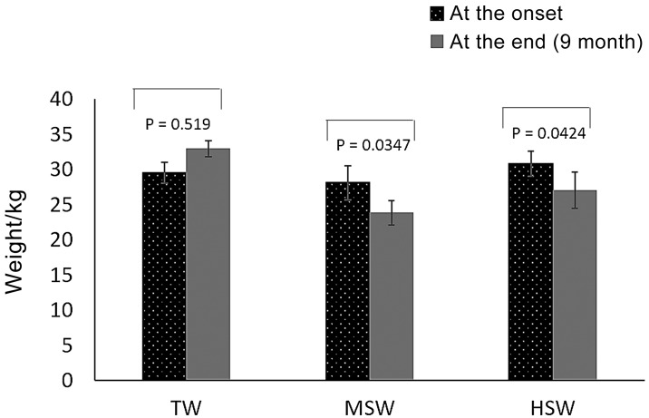
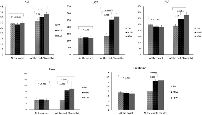
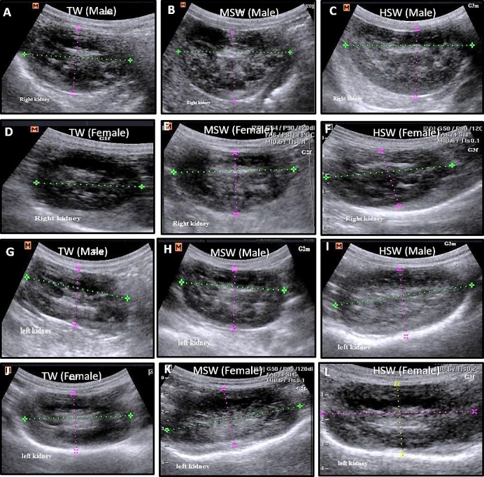
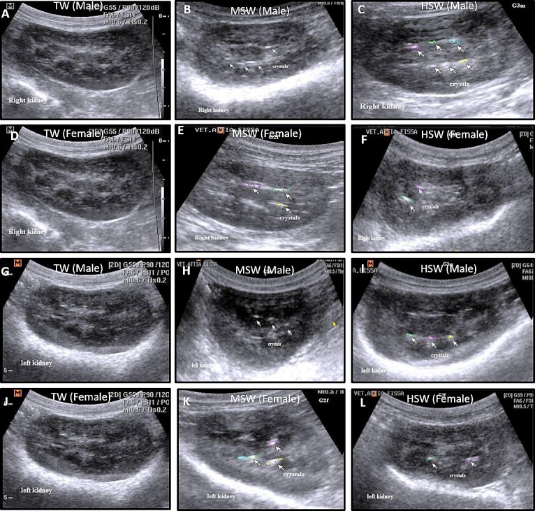
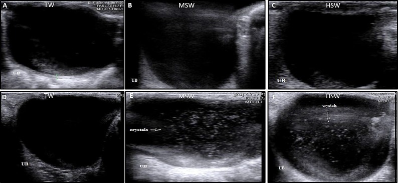
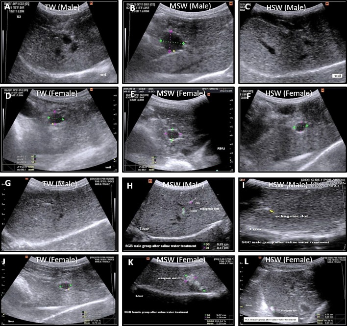
The objective of this study was to evaluate the impact of varying degrees of water salinity on the ultrasonographical and histopathological appearance of the liver and kidneys in Barki sheep. Thirty Barki sheep (initial weight, 29.48 ± 0.81 kg) were allocated into three groups (n=10 per group) based on the type of drinking water for 9 months: the tap water (TW) group (350 ppm total dissolved solids [TDS]); the moderate saline water (MSW) group (4,557 ppm TDS); and the high saline water (HSW) group (8,934 ppm TDS). After 9 months, the body weight was significantly decreased in sheep subjected to MSW (P=0.0347) and HSW (P=0.0424). Alanine aminotransferase, aspartate aminotransferase, alkaline phosphatase, urea, and creatinine were significantly increased (P<0.05) in sheep subjected to MSW and HSW. Ultrasonographic examination of the right and left kidneys revealed an increased length of both kidneys with crystal formation, particularly in male sheep. Ultrasonographic examination of the liver showed hyperechogenic dots varying in size and number between males and females. Histopathological examination of kidney revealed significant changes in both MSW and HSW groups such as hyaline matrix formation, atrophied glomerular tufts, and intramedullary congestion. Histopathological examination of the liver revealed slight fatty liver changes, slight fibrosis around the bile duct, massive inflammatory cell infiltration and vacuolar changes of hepatocytes in both MSW and HSW groups. In conclusion, water salinity negatively affects the body weight, liver and kidney appearance of Barki sheep and thus sheep production.
Keywords: histopathology; kidney; liver; sheep; ultrasonography.
Figures
Similar articles
-
The effects of saline water consumption on sperm parameters, testicular histopathology, hormonal and antioxidants concentrations in Barki Rams.BMC Vet Res. 2024 May 23;20(1):219. doi: 10.1186/s12917-024-04047-2. BMC Vet Res. 2024. PMID: 38778406 Free PMC article.
-
NTP Toxicology and Carcinogenesis Studies of 4,4'-Thiobis(6- t -butyl- m -cresol) (CAS No. 96-69-5) in F344/N Rats and B6C3F1 Mice (Feed Studies).Natl Toxicol Program Tech Rep Ser. 1994 Dec;435:1-288. Natl Toxicol Program Tech Rep Ser. 1994. PMID: 12595928
-
NTP Toxicology and Carcinogenesis Studies of Pyridine (CAS No. 110-86-1) in F344/N Rats, Wistar Rats, and B6C3F1 Mice (Drinking Water Studies).Natl Toxicol Program Tech Rep Ser. 2000 Mar;470:1-330. Natl Toxicol Program Tech Rep Ser. 2000. PMID: 12579203
-
Toxicology and carcinogenesis studies of milk thistle extract (CAS No. 84604-20-6) in F344/N rats and B6C3F1 mice (Feed Studies).Natl Toxicol Program Tech Rep Ser. 2011 May;(565):1-177. Natl Toxicol Program Tech Rep Ser. 2011. PMID: 21685957 Review.
-
Toxicology and carcinogenesis studies of androstenedione (CAS No. 63-05-8) in F344/N rats and B6C3F1 mice (gavage studies).Natl Toxicol Program Tech Rep Ser. 2010 Sep;(560):1, 7-31,33-171 passim. Natl Toxicol Program Tech Rep Ser. 2010. PMID: 21037592 Review.
Cited by
-
Blood biochemical parameters of Murrah buffalo (Bubalus bubalis) reared in the high salinity area of Bangladesh.J Adv Vet Anim Res. 2022 Dec 31;9(4):736-742. doi: 10.5455/javar.2022.i643. eCollection 2022 Dec. J Adv Vet Anim Res. 2022. PMID: 36714502 Free PMC article.
-
Ultrasound imaging of the spleen, liver, and kidneys in healthy hair sheep: a pilot study.Trop Anim Health Prod. 2025 Mar 14;57(2):119. doi: 10.1007/s11250-025-04375-x. Trop Anim Health Prod. 2025. PMID: 40085310
-
Physiological, hematological, and biochemical responses in Hararghe-highland lamb subjected to water salinity levels of Lake Basaka in a semiarid area of Ethiopia.Heliyon. 2022 Dec 22;8(12):e12616. doi: 10.1016/j.heliyon.2022.e12616. eCollection 2022 Dec. Heliyon. 2022. PMID: 36619434 Free PMC article.
-
Effects of Salinity Levels of Drinking Water on Water Intake and Loss, Feed Utilization, Body Weight, Thermoregulatory Traits, and Blood Constituents in Growing and Mature Blackhead Ogaden Sheep and Somali Goats.Animals (Basel). 2024 May 25;14(11):1565. doi: 10.3390/ani14111565. Animals (Basel). 2024. PMID: 38891612 Free PMC article.
-
Impact of drinking of saline water on hemato-biochemical parameters of Black Bengal goats in the selected areas of Bangladesh.Saudi J Biol Sci. 2022 Oct;29(10):103397. doi: 10.1016/j.sjbs.2022.103397. Epub 2022 Jul 25. Saudi J Biol Sci. 2022. PMID: 35991851 Free PMC article.
References
-
- Assad F., El-Sherif M. A.2002. Effect of drinking saline water and feed shortage on adaptive responses of sheep and camels. Small Rumin. Res. 45: 279–290. doi: 10.1016/S0921-4488(02)00083-4 - DOI
-
- Attia-Ismail S. A., Abdo A. R., Asker A. R. T.2008. Effect of salinity level in drinking water on feed intake, nutrient utilization, water intake and turnover and rumen function in sheep and goats. Egypt. J. Sheep Goats Sci. 3: 77–94.
-
- Bancroft J. D., Gamble M.2008. Theory and practice of histological techniques book. Churchill Livingstone, New York /Elsevier.
MeSH terms
Substances
LinkOut - more resources
Full Text Sources
Other Literature Sources
Medical
Miscellaneous